University of Groningen. Oral sequelae resulting from head and neck radiotherapy Jansma, Johan
|
|
|
- Jeroen Geerts
- 4 jaren geleden
- Aantal bezoeken:
Transcriptie
1 University of Groningen Oral sequelae resulting from head and neck radiotherapy Jansma, Johan IMPORTANT NOTE: You are advised to consult the publisher's version (publisher's PDF) if you wish to cite from it. Please check the document version below. Document Version Publisher's PDF, also known as Version of record Publication date: 1991 Link to publication in University of Groningen/UMCG research database Citation for published version (APA): Jansma, J. (1991). Oral sequelae resulting from head and neck radiotherapy: Course, prevention and management of radiation caries and other oral complications. [S.l.]: [S.n.]. Copyright Other than for strictly personal use, it is not permitted to download or to forward/distribute the text or part of it without the consent of the author(s) and/or copyright holder(s), unless the work is under an open content license (like Creative Commons). Take-down policy If you believe that this document breaches copyright please contact us providing details, and we will remove access to the work immediately and investigate your claim. Downloaded from the University of Groningen/UMCG research database (Pure): For technical reasons the number of authors shown on this cover page is limited to 10 maximum. Download date:


2 Oral sequelae resulting from head and neck radiotherapy Course, prevention and management of radiation caries and other oral complications Johan Jansma
3 ORAL SEQUELAE RESULTING FROM HEAD AND NECK RADIOTHERAPY Course, prevention and management of radiation caries and other oral complications
4 Cover: T. Jansma & E. G. C. van Ommen Typesetting: STYX Publications
5 STELLING EN behorend bij het proefschrift ORAL SEQUEL AE RESULTING FROM HEAD AND NECK RADIOTHERAPY Course, prevention and management of radiation caries and other oral complications Tandglazuur bestraald met een therapeutische dosis ioniserende stralen, ondervindt hiervan geen schadelijke gevolgen. Dit proefschrift 2 De in de literatuur geadviseerde dagelijkse applicatie van een fluoride-gel bij patienten bestraald op het hoofd-halsgebied kan, mits gecombineerd met een goede mondhygiene, worden teruggebracht tot eenmaal per twee dagen. Dit proefschrift 3 Bij de bestralingsbehandeling van patienten met hoofd-halstumoren dient men te beschikken over een goed opgeleid tandheelkundig team. 4 Hyposialie wordt door veel clinici onderschat. 5 Een speekselsubstituut op maat bestaat niet. (Levine et al. J Dent Res 1987;66: ) 6 Het onderzoek van Franzen et al. naar de effecten van gefractioneerde bestraling op de morfologie en functie van speekselklierweefsel gaat voorbij aan de huidige inzichten in de mechanismen van stralingsschade aan dat weefsel. (Franzen et al. Lab Invest 1991;64: ) 7 Een Abbe-plastiek moet bij schisispatienten niet worden toegepast indien er geen normale intermaxillaire relatie bestaat. 8 Het is te hopen dat er na de stormachtige ontwikkeling die de orale implantologie heeft doorgemaakt geen toekomst is weggelegd voor de orale explantologie. 9 Het verwijderen van een verstandskies in de onderkaak zonder gebruikmaking van een adequate rontgenfoto getuigt van weinig gevoel voor de onderlip.
6 10 De relatie tussen de concentratie N-acetyi-L-aspartaat in bepaalde hersengebieden en de ernst van dementie bij de ziekte van Alzheimer is aan kritiek onderhevig. (Kwo-On-Yuen et al. Soc Magn Res Med 1991;1:429) II Diabetische retinopathie is aileen goed te beoordelen indien gespiegeld wordt in mydriasis. 12 In het kader van infectie-preventie, waarvan men zich steeds meer bewust is geworden door de AIDS problematiek, dienen kappers voor iedere klant een nieuw scheermes te gebruiken. 13 De term kijkoperatie dient te worden gereserveerd voor die ingrepen waarbij de assistent in opleiding slechts mag toekijken. 14 Veel politie-agenten en militairen ontlenen een deel van hun gezag aan hun snor. Groningen, 13 november 1991 Johan Jansma
7 RIJKSUNIVERSITEIT GRONINGEN ORAL SEQUELAE RESULTING FROM HEAD AND NECK RADIOTHERAPY Course, prevention and management of radiation caries and other oral complications PROEFSCHRIFr ter verkrijging van bet doctoraat in de Geneeskunde aan de Rijksuniversiteit Groningen op gezag van de Rector Magnificus Dr. S. K. Kuipers in bet openbaar te verdedigen op woensdag 13 november 1991 des namiddags te 2.45 uur precies door JOHAN JANSMA geboren op 24 januari 1962 te Leeuwarden DRUKKERIJ VAN DENDEREN B.V. Groningen 1991
8 Promotores Prof. Dr. E. J. 's-gravenmade Prof. Dr. A. K. Panders Prof. Dr. A. Vermey, FACS Referent Dr. A. Vissink Promotiecommissie Prof. Dr. G. Boering Prof. Dr. D. H. Retief Prof. Dr. B. G. Szabo Paranimfen Drs. A. G. Becking Drs. R. J. Bun
9 Aan mijn ouders Voor Dionne
10 ISBN J. Jansma, Groningen, 1991 Parts of this study were financially supported by: Koningin Wilhelmina Fonds (grant no. 84-4) Praeventiefonds (grant no ) Contributions to the printing costs of this thesis were made by: Stichting Het Scholten-Cordes Fonds Dr. L.A. Burna Stichting Pharmachemie BY
11 CONTENTS VOORWOORD 1 INTRODUCfiON AND AIMS OF THE INVESTIGATION 2 REVIEW OF THE LITERATURE Introduction Radiation treatment Oral consequences of head and neck radiotherapy Oral mucosa Salivary glands Dentition Periodontium Bone Taste Muscles and joints Pattern of complaints Prevention and treatment of oral sequelae resulting from head and neck radiotherapy Mucositis Hyposalivation Radiation caries Periodontal disease Osteoradionecrosis Taste loss Trismus 3 RADIATION CARIES: IN VITRO AND IN SITU EXPERIMENTS The effect of X-ray irradiation on the demineralization of bovine dental enamel The effect of X-ray irradiation on the permeability of bovine dental enamel A model to investigate xerostomia-related dental caries A SEM study of natural and induced radiation caries In situ study on the prevention of radiation caries 4 A SURVEY OF THE PREVENTION AND TREATMENT REGIMENS OF ORAL SEQUELAE RESULTING FROM HEAD AND NECK RADIOTHERAPY USED IN DUTCH RADIOTHERAPY INSTITUTES vii
12 5 PROTOCOL FOR THE PREVENTION AND TREATMENT OF ORAL SEQUEL AE RESULTING FROM HEAD AND NECK RADIOTHERAPY Introduction Prevention and treatment of oral sequelae Pre-irradiation patient management Physical and radiographic examination Treatment and prophylaxis Initiation of a new preventive regimen Nutritional instructions Patient management during radiation treatment Post-irradiation patient management Oral mucosa Oral dryness Dentition Trismus prevention Nutritional counseling Epilogue SUMMARY SAMENVATTING APPENDIX: QUESTIONNAIRE CURRICULUM VITAE viii
13 VOORWOORD Toen ik vijf jaar geleden met dit onderzoek begon en bet nog niet vast stond dat bet tot een proefschrift zou leiden, had ik er geen idee van dat ik met zeer vee! verschillende mensen van zeer verschillende afdelingen op uiterst plezierige wijze zou gaan samenwerken. Ik kijk dan ook terug op een boeiende en leerzame periode die ik beslist niet had willen missen. Op deze plaats wil ik graag al diegenen bedanken, die op enigerlei wijze hebben bijgedragen aan bet tot stand komen van dit proefschrift. Een aantal personen wil ik met name noemen. Prof. Dr. E. J. 's-gravenmade, hooggeachte promotor, beste professor. U bent degene geweest die mijn interesse heeft gewekt voor bet verrichten van een promotie-onderzoek. De positief kritische en altijd bijzonder stimulerende wijze waarop U dit werk vanaf bet begin hebt begeleid en de wijze waarop U mij wegwijs maakte in 'onderzoeksland' heb ik zeer gewaardeerd. Mijn dank daarvoor is groot. Prof. Dr. A. K. Panders, hooggeachte promotor, beste Arend. Jouw grote klinische kennis en ervaring waren onontbeerlijk voor bet klinische dee! van bet onderzoek. Je commentaren en onze discussies heb ik als zeer waardevol ervaren en hoop ze, ook binnen andere richtingen van ons vak, nog vaak met je over te doen. Prof. Dr. A. Vermey, hooggeachte promotor, beste professor. Ik ben blij dat U, als nestor van de preventieve zorg bij hoofd-halsbestralings patienten in Groningen, betrokken was bij mijn onderzoek. U bent reeds vaak geroemd vanwege de snelheid waarmee U manuscripten op kundige wijze van commentaar voorziet en ik sluit mij in dat opzicht dan ook graag bij vorige promovendi aan. Bedankt voor de prettige samenwerking. Dr. A. Vissink, zeer geleerde referent, beste Arjan. Een ieder die jou kent, hoef ik bet niet uit te leggen en een ieder die jou niet kent, zal bet niet direct begrijpen, maar een betere begeleider dan jij bestaat er niet. Je was zeer betrokken bij bet onderwerp en het onderzoek met zowel raad als daad. Ik heb erg vee! van je geleerd en altijd uiterst plezierig en vriendschappelijk met je samengewerkt en hoop dat ook in de toekomst te blijven doen. Prof. Dr. G. Boering, beste professor. Ik ben U zeer dankbaar dat U mij de mogelijkheid hebt geboden om naast mijn opleiding tot kaakchirurg mijn onderzoek voort te zetten. U was altijd zeer gelnteresseerd in mijn vorderingen. lk beschouw bet als een eer, dat U bereid was zitting te nemen in de promotiecommissie. Prof. Dr. D. H. Retief, beste professor. Ek is U groot dank verskuldig vir die samenwerking met U gedurende die eerste jaar van die navorsingsprojek asook vir die vinnige beoordeling van hierdie tesis. Ek beskou dit as 'n groot eer dat U bij die promosie teenwoordig sal wees. Baie, baie dankie. Prof. Dr. B. G. Szabo, beste professor. Graag wil ik U bedanken voor Uw welwillendheid om dee! uit te maken van de promotiecommissie en voor de tijd en aandacht die U aan mijn proefschrift hebt willen besteden. IX
14 Dr. W. L. Jongebloed, beste Wim. Ik benjou, als expert op het gebied van caries in de hoogste vergroting, vee! dank veschuldigd voor de grote vakkundigheid waarmee je de vele scanning-elektronenmicroscopische opnamen hebt gemaakt en voor je inbreng in de discussies over wat er allemaal op te zien was. Prof. Dr. B. Lowenberg van het Rotterdams Radio-Therapeutisch Instituut/ Daniel den Hoed Kliniek dank ik hartelijk voor de gelegenheid mij geboden om gedurende een jaar verbonden te zijn geweest aan genoemd instituut. Drs. L. L. Visch, beste Leo. Onze contacten in de beginfase van het onderzoek hebben in belangrijke mate bijgedragen tot wat het onderzoek uiteindelijk is geworden. Bedankt daarvoor. Prof. Dr. F. C. M. Driessens, Dr. J. M.P. M. Borggreven en Dhr. R.Gorissen van de afdelingen Tandheelkundige Materialen en Biochemie M.W. van de Katholieke Universiteit Nijmegen. Hartelijk dank voor de goede en efficiente samenwerking bij de diffusie- en impedantie-experimenten en voor de hulp bij het op schrift stellen van deze, wat mij betreft, zeer ingewikkelde materie. Dr. J. A K. M. Buskes, beste Hans. Hartelijk dank voor je begeleiding tijdens mijn eerste stappen 'in' jouw 'constant composition' apparaat en voor je hulp bij mijn eerste geschriften. Dr. V. Fidler wil ik danken voor het uitvoeren van de statistische analyses bij het caries onderzoek, waarvan U altijd zei dat het sneller was om ze zelf uit te voeren, dan om ze aan mij uit te leggen. Prof. Dr. W. G. Perdok, beste professor. Hartelijk dank voor het uitvoeren van het rontgendiffractie onderzoek en de interpretatie van de gegevens. Mw.l. Retief. Baie dankie vir die nougesetheid waarmee U die fluoridebepalings uitgevoer het. Drs. D. M. Mehta en Dhr. A A Canrinus wil ik danken voor hun hulp bij het bestralen van de vele glazuurpreparaten, beter bekend als 'koeietanden' en de adviezen daaromtrent. Dr. E. de Josselin de Jong, beste Elbert. Bedankt voor je microradiografische adviezen in de ontwikkelingfase van het cariesmodel. Prof. Dr. J. Arends en Prof. Dr. J.J. ten Bosch. In de beginfase van mijn onderzoek heb ik veelvuldig gebruik mogen maken van het goed geoutilleerde laboratorium voor Materia Technica. Ik ben U heiden daarvoor zeer erkentelijk. Dr. J. Bouma, beste Jelte. Jij hebt me wegwijs gemaakt in de wereld van het interview en de vragenlijst. Ik hoop dat je mij in de toekomst nog eens vaker iets wijs zult willen maken. Drs. H. J. Guchelaar, beste Henk-Jan. Dank voor je hulp bij de ontwikkeling en verfijning van de samenstelling van de fluoride gel. Dr. F. K. L. Spijkervet en Dr. J. L. N. Roodenburg, beste Fred en Jan. Hartelijk dank voor jullie expertise bij de ontwikkeling van het protocol. Fred wil ik in het bijzonder bedanken voor onze vele vruchtbare discussies die, hoewel vaak gramnegatief getint, voor mij een zeer positieve herinnering vormen. De heren M. Hummel en A Wietsma, beste Martin en Anne. Dankzij jullie vakmanschap leken mijn glazuurhouders wei van zwitserse makelij. Reuze bedankt. X
15 Mw. L. E. Noordhof wit ik graag bedanken voor de hulp bij het opsporen van de vele, soms haast onvindbare, literatuur. Dr. J. K. Tidwell, dear John. Thanks for the many fruitful discussions we had during the Arnhem period and for correcting parts of the English text. Mw. M.A. de Jong, beste Mirabelle. Bedankt voor je correcties van mijn gebruik van ons Nederlands. De heren P. van der Sijde, D. Huizinga, H. R. A. Meiborg, R. L. Dijkstra en H. R. Luurtsema wit ik bedanken voor de vele fraaie foto's die zij in de loop der jaren voor mij hebben gemaakt en waarvan een deel nu mijn proefschrift siert. De heren H. Flanderijn en E. G. C. van Ommen, beste Henk en Erik. Zeer bedankt voor jullie professionele tekenwerk en voor het goede contact dat we er altijd over hadden. Drs. G. Steensma en Drs. J. W. Braams, beste Gauke en Jan-Willem. Bedankt voor het kritisch doorlezen van de zetproeven. Dankzij jullie staan de puntjes nu op de i. Mw. G. Boezerooy-Nobach, Mw. K. Wolthuis en Mw. E. van Drooge, beste Gerda, Karin en Ellen. Bedankt voor jullie secretari(he en morele ondersteuning. De medewerkers van aile Nederlandse afdelingen voor Radiotherapie en hun tandheelkundige teams wit ik graag bedanken voor hun spontane medewerking aan het inventarisatie onderzoek. Alle medewerkers, waarvan enkele inmiddels ex-medewerkers, van de afdeling Mondziekten, Kaakchirurgie en Bijzondere Tandheelkunde, die hebben bijgedragen aan een prettige werksfeer en be grip toonden wanneer ik tijdens de experimenten en in de afrondingsfase af en toe eens verstek moest Iaten gaan, wil ik hartelijk bedanken. Aile patienten die geheel belangeloos hun medewerking verleenden aan mijn experimenten en bij wie ik vaak wekelijks zeer gastvrij werd onthaald, mijn hartelijke dank. Mijn ouders, jullie dank ik dat jullie mij in de gelegenheid hebben gesteld om een universitaire opleiding te volgen en vooral voor de warme belangstelling en interesse die jullie steeds voor mijn bezigheden hebben getoond. Aan jullie draag ik daarom mijn proefschrift op. Dionne. Het tot stand komen van dit proefschrift is niet in de laatste plaats ook jouw verdienste. Jij was mijn dagelijkse inspiratiebron en mijn belangrijkste coauteur. Ik ben je zeer dankbaar voor je opgewektheid, je steun en al je geduld. The research was carried out at the Department of Oral and Maxillofacial Surgery of the University Hospital Groningen in cooperation with the Laboratory for Materia Technica, the Department of Radiotherapy and the Laboratory for Histology and Cell Biology of the University of Groningen; the Daniel den Hoed Kliniek!Rotterdam Radiotherapeutic Institute; the Departments of Oral Biomaterials and Biochemistry of the University of Nijmegen and the Institute of Dental Research of the University of Alabama, u.s.a. XI
16
17 Chapter 1 INTRODUCTION AND AIMS OF THE INVESTIGATION INTRODUCTION Malignant tumors of the head and neck comprise all malignant tumors located above the level of the clavicles with the exception of those originating from the central nervous system. These tumors can be grouped according to the anatomic location and histology of the involved tissues and include: facial skin and lips, upper aero-digestive tract (nasal cavity, paranasal sinuses, oral cavity, pharynx, cervical oesophagus, larynx, trachea), salivary glands, thyroid and parathyroid glands, bone and soft tissues, orbits, and lymph nodes. Similarity in etiology, patterns of metastasis, diagnostics and treatment justify these malignant neoplasms to be put together in one group: head and neck cancer. The incidence of head and neck cancer (skin cancer excluded) in Western Europe and the U.S.A. is about 23/100,000 inhabitants, i.e. about 8% of all malignant tumors.1 The tumors addressed in this thesis are especially those malignant tumors originating from the upper aero-digestive tract. Malignant neoplasms in the head and neck region are often treated with a combination of surgery and radiotherapy, while malignant lymphomas in this area are usually treated with radiotherapy and/or chemotherapy.2 In addition to anti-tumor effects, radiation also induces damage in normal tissues, irrespective of individual treatment plans and in spite of the improved tissue-sparing properties of modern radiation techniques. In patients with head and neck cancer, radiation treatment often involves the tissues of the oral cavity and the major and minor salivary glands as well as the jaws, either because of the location of the primary tumor or of the lymph node metastases. Radiation injury of salivary glands, oral mucosa and jaws may lead to early or late occurring oral sequelae like xerostomia,3 taste loss,4 irradiation mucositis,4 5 radiation caries,6 7 soft-tissue necrosis and osteoradionecrosis of the jaw bone.8-10 These sequelae form a heavy burden for these patients, may cause a lot of pain and distress during and after radiotherapy and may become dose-limiting. Similar oral sequelae (e.g. mucositis, hyposalivation) can be observed during a course of chemotherapy, but they have a more temporary character There are many indications that oral sequelae resulting from head and neck radiotherapy can be prevented or reduced in severity As the prognosis for localized head and neck cancer and malignant lymphomas is relatively favorable, 5-year survival rates of 50% or more have been reported,21 22 the dental and oral status of the patient should be considered with great care. Especially in cases of tumor control, irradiation-induced changes in oral and adjacent structures are major factors determining the patient's quality of life. 1
18 Concerning the prevention of radiation caries, only a few of the approaches reported in the literature are based on fundamental research These caries prophylactic regimens are inconvenient for the patient because of the lifelong need for daily fluoride applications. This implies a risk of compliance failure leading to increased caries activity. Preservation of a healthy dentition is a matter of increasing significance since the number of (aged) dentulous patients and the dental mindedness in developed countries is increasing considerably. Optimization of radiation caries prevention as one of the main missing links in a proper overall protocol for the prevention and treatment of oral sequelae resulting from head and neck radiotherapy is therefore a major subject of this thesis. AIMS OF THE INVESTIGATION The general aim of this thesis is to study the course and prevention of radiation caries and to propose an overall protocol for the prevention and treatment of oral sequelae resulting from head and neck radiotherapy. The specific aims are: - to review the literature regarding the effects of ionizing irradiation on nonnal tissues in the head and neck region, the resulting sequelae and their prevention and treatment, specifically to develop an overall preventive protocol (chapter 2). Radiation treatment in the head and neck region may cause many changes in normal tissues resulting in e.g. mucositis, hyposalivation and radiation caries. Knowledge of these sequelae as well as their prevention and treatment is essential to develop a proper preventive protocol (chapter 5). - to study the course and prevention of radiation caries (chapter 3). Irradiation-induced hyposalivation, the resulting changes in the composition of saliva and oral flora and the altered dietary habits are all agreed upon indirect factors contributing to an increased caries susceptibility after radiotherapy. The direct effect of ionizing irradiation on the (in )organic components of dental enamel is unclear. Furthermore, there is no proper model known in the literature suitable to study the development and prevention of radiation caries. The following in vitro and in situ studies were performed: - studies on the direct effects of ionizing irradiation on the acid solubility and permeability of dental enamel; - development of an in situ model for studying the onset, progression and prevention of radiation caries as a function of time; - comparison of the initiation and progression of induced radiation caries with those of natural radiation caries; - study on the prevention of radiation caries by evaluating the effects of different fluoride concentrations and application procedures. - to survey the prevention and treatment regimens of oral sequelae resulting from head and neck radiotherapy applied in all radiotherapy institutes in the Netherlands and to evaluate the differences in these regimens (chapter 4). 2
19 Most prevention procedures described in the literature are based on clinical experience. The result is a great diversity in policies, which may lead to a variety in the preventive approach in daily practice. To study this assumption a survey of the preventive regimens applied in all Dutch radiotherapy institutes was performed. A survey of these regimens is needed to determine what caries prophylactic methods and other preventive measures are used in a clinical setting as well as to assess the need for the development of an overall protocol for the prevention and treatment of oral complications of head and neck radiotherapy. This survey was not intended to evaluate the effects of the various regimens in the patient situation. to develop an overall protocol for the prevention and treatment of oral sequelae resulting from head and neck radiotherapy (chapter 5). When covering the subject of radiation caries it is difficult to look at this side effect as a single entity without taking into account other side effects. Hyposalivation, mucositis and trismus all directly interfere with the oral status, the well-being, the degree of oral hygiene and the dietary habits of the patient and emphasize the need for an overall prevention and treatment protocol. Furthermore, such a protocol can be a major tool in the prevention of acute exacerbation of foci of infection and the prevention of osteoradionecrosis. A scientific basis for the development of a feasible overall protocol for the prevention and treatment of oral sequelae resulting from head and neck radiotherapy is formed by the hyposalivation studies of Vissink, 18 the mucositis studies of Spijkervet20 and the radiation caries studies described in this thesis in combination with data derived from the literature (chapter 2). REFERENCES Vermey A, Oldhoff J, Panders AK. Hoofd-hals oncologie. In: de Boer J, Derom F, Gruwez JA, Kuijer PJ, den Otter G, Zwaveling A, eds. Leerboek chirurgie 3rd ed. Bohn, Scheltema & Holkema, Utrecht/ Antwerpen 1988, Al-Sarraf M. Chemotherapeutic management of head and neck neoplasia. In: Peterson DE, Elias EG, Sonis ST, eds. Head and neck management of the cancer patient. Martinus Nijhoff, Boston 1986, Dreizen S, Brown LR, HandlerS, Levy BM. Radiation induced xerostomia in cancer patients. Effect on salivary and serum electrolytes. Cancer 1976;38: Beumer J, Curtis T, Harrison RE. Radiation therapy of the oral cavity: Sequelae and management. Part 1. Head Neck Surg 1979;1: Lockhart PB. Oral complications of radiation therapy. In: Peterson DE, Elias EG, Sonis ST, eds. Head and neck management of the cancer patient. Martin us Nijhoff, Boston 1986, Frank RM, Herdly J, Philippe E. Acquired dental defects and salivary gland lesions after irradiation for carcinoma. JAm Dent Assoc 1965;70: Karmiol M, Walsh RF. Dental caries after radiotherapy of the oral regions. JAm DentAssoc 1975;91: Murray CG, Herson J, Daly TE, Zimmerman S. Radiation necrosis of the mandible: a 10-year study. Part I. Factors influencing the onset of necrosis. Int J Radiat Oncol Bioi Phys 1980;6: Murray CG, Herson J, Daly TE, Zimmerman S. Radiation necrosis of the mandible: a 10-year study. Part II. Dental factors, onset, duration and management of necrosis. Int J Radiat Oncol Bioi Phys 1980;6: I 0 Marx RE. Osteoradionecrosis. A new concept of its pathophysiology. J Oral Maxillofac Surg 1983;41:
20 11 Sonis ST, Sonis AL, Lieberman A. Oral complications in patients receiving treatment for malignancies other than the head and neck. JAm Dent Assoc 1978;97: Dreizen S, McCredie KB, Bodey GP, Keating MJ. Quantitative analysis of the oral complications of antileukemia chemotherapy. Oral Surg Oral Med Oral Pathol 1986;62: Daly TE, Drane JB, MacComb WS. Management of problems of the teeth and jaw in patients undergoing irradiation. Am J Surg 1972;124: Keys HM, McCasland JP. Techniques and results of a comprehensive dental care program in head and neck cancer patients. Int J Radiat Oncol Bioi Phys 1976;1: Dreizen S, Brown LR, Daly TE, Drane JB. Prevention of xerostomia-related dental caries in irradiated cancer patients. J Dent Res 1977;56: Horiot JC, Schraub S, Bone MC, Bain Y, Ramadier J, Chaplain G, Nabid N, Thevenot B, Bransfield D. Dental preservation in patients irradiated for head and neck tumours: A 10-year experience with topical fluoride and a randomized trial between two fluoridation methods. Radiother Oncol 1983;1: Toljanic JA, Saunders VW. Radiation therapy and management of the irradiated patient. J Prosthet Dent 1984;52: Vissink A. Xerostomia. Development, properties and application of a mucin-containing saliva substitute; thesis Groningen, Wright WE, Haller M, Harlow SA, Pizzo PA. An oral disease program for patients receiving radiation and chemotherapy. JAm Dent Assoc 1985;110: Spijkervet FKL. Irradiation mucositis and oral flora. Reduction of mucositis by selective elimination of oral flora; thesis Groningen, Hakulinen T, Pukkala E, Hakama M, Lehtonen M, Saxn, E, Teppo L. Survival of cancer patients in Finland in Ann Clin Res 1981;13:Suppl 31, Sonis ST. Cancer, its complications, and the head and neck. In: Peterson DE, Elias EG, Sonis ST, eds. Head and neck management of the cancer patient. Martin us Nijhoff, Boston 1986,
21 Chapter 2 REVIEW OF THE LITERATURE INTRODUCTION In addition to anti-tumor effects, ionizing irradiation causes damage in normal tissues located in the field of radiation. The oral complications of radiotherapy in the head and neck region are the result of the deleterious effects of irradiation on e.g. salivary glands, oral mucosa, bone, dentition, masticatory musculature and temporomandibular joints (TMJ). The clinical consequences of radiation treatment include mucositis, hyposalivation, taste loss, osteoradionecrosis, radiation caries and trismus. Mucositis and taste loss are reversible consequences that usually subside early post-irradiation while hyposalivation is normally an irreversible effect. Furthermore, the risk of developing radiation caries and osteoradionecrosis are life-long threats. All these consequences form a heavy burden for the patients and have a tremendous impact on their quality of life during and after radiotherapy. In this chapter the role of radiation treatment in head and neck oncology, the irradiation-induced changes in normal oral tissues, the resulting clinical consequences, and the possibilities for prevention and treatment of these consequences are reviewed. The irradiation-related changes in the oral mucosa, salivary glands, dentition, periodontium, bone, muscles and joints are described in that order. RADIATION TREATMENT Radiation treatment plays an important role in the management of head and neck cancer. According to Rothwell approximately 50% of all new cases of invasive head and neck cancer will need radiation therapy as a primary treatment, as palliation, or as an adjunct to surgery or chemotherapy.1 Low energy, superficial ( kv) and orthovoltage ( kv) irradiation, is used in the treatment of skin and other superficial tumors, whereas high energy external beam irradiation (500 kv- 20 MY) including cobalt-60 irradiation and megavoltage irradiation produced by linear accelerators (8-20 MY photons, 4-20 MeV electrons) and internal irradiation are used in the treatment of deeper located malignancies.2 Important advantages of high energy irradiation as compared to low energy irradiation in the treatment of head and neck cancer are: reduced absorption in bone, less damage to the skin and reduced scatter of radiation into other body tissues. The result is a decrease of the incidence and severity of head and neck complications.3 The advantage of internal radiotherapy (interstitial or intracavitary with radium needles, radon seeds and other radioactive elements such as cesium and iridium) is that 5

22 0\ o::::> a Rs 28 c,.., - f' t ',\. + '\ b n:.::> / <-.. l f \ \ c d 1%\ e Fig. 1 Common fields of radiation therapy for head and neck tumors are shown schematically by solid lines. Dotted lines indicate fields when increased dosages are used. A Parotid field. 8 Antrum field. C Oropharynx field. D Floor of mouth field. E Mantle fields for Hodgkin disease. F Fields for lymphatics of the neck. 80 f
23 there is a high irradiation dose directed to the tumor without a high dose to the surrounding tissues. Internal irradiation may be used for the total treatment or in combination with external irradiation.4 The radiation dose needed for the treatment of cancer is based on location and type of malignancy, and whether or not radiotherapy will be used solely or as a preor postsurgical treatment modality. Most patients with head and neck carcinomas receive between 50 and 70 Gy (1 Gy = 1 J/kg = 100 rad) as a curative dose. This dose is usually given over a five to seven week period, once a day, five days a week, with a daily tumor dose of about 2 Gy. The advantages of fractionated irradiation in contrast with single dose treatment are that time is allowed for healthy tissues to repair and repopulate and for the tumor to 'shrink' slowly, enabling reoxygenation and redistribution of the hypoxic tumor cell fraction and therefore more efficient treatment. 4 In case of preoperative irradiation or irradiation for malignant lymphomas usually lower total doses are used.5 6 Because of the location of the primary tumor and regional lymph nodes, the oral cavity, salivary glands and jaws of most head and neck cancer patients are in the field of radiation (Fig. 1 ). As a result, unwanted radiation induced changes will occur in these tissues. An important limiting factor in tumor irradiation is the radiation dose that adjacent normal tissues can tolerate. Tissues with rapid turnover rates show acute reactions to irradiation (early effects), while in tissues with slower turnover rates damage may not become evident for months or years after therapy (late effects ).1 ORAL CONSEQUENCES OF HEAD AND NECK RADIOTHERAPY Oral mucosa The damage to the oral mucosa in the path of radiation depends upon the total irradiation dose, volume of irradiated tissue, fractionation and type of ionizing irradiation.4 8 As mucosal cells turn over rapidly, they have a low resistance to radiation. Oral side effects following therapeutic irradiation therefore develop quickly.9 Mucositis induced by irradiation is defined as the reactive inflammatory-like process of the oropharyngeal mucous membrane during therapeutic head and neck irradiation. Irradiation mucositis is an inevitable but transient side effect It is an integral part of the radiation therapy in terms of morbidity. The early radiation reaction causes local discomfort as well as difficulties in drinking, eating, swallowing and speech. Therefore it can give rise to nutritional problems and in severe cases nasogastric feeding may become necessary, which is very uncomfortable Severe mucositis may necessitate an interruption of the course of radiation treatment and thus can serve as a dose-limiting factor. 1 3 Such interruptions must be 7
24 prevented because they may result in prolongation of treatment and reduction in therapeutic effect Various signs of mucositis may emerge during radiotherapy The first clinical signs of mucositis occur at the end of the first week of a conventional six week radiation protocol (daily dose of 2 Gy, five times a week). There is no consensus regarding what is the first sign of mucositis. Some authors describe a white discoloration of the oral mucosa, which is an expression of hyperkeratinization as the first symptom, followed by or in combination with erythema Others consider erythema, due to vascular dilatation (hyperaemia) or obstructive changes in arterioles, to be the first reaction As radiation therapy progresses, superficial cells are no longer replaced because irradiation-induced cell loss in the basal cell layer exceeds the proliferation of new cells, and the mucosa becomes thin, red and friable. 20 Vascular dilatation and edema of the submucosa lead to further weakening ofthe oral mucosa and can provoke an epithelial breakdown.3 After about three weeks of irradiation more severe symptoms of mucositis such as the formation of pseudomembranes and ulcerations may appear Some authors consider pseudomembranes to be ulcers covered by fibrinous exudate.3 21 Others suggest pseudomembranes to be related to yeast stomatitis19 or to colonization of the oral cavity with gram-negative bacilli. 17 Mucosal ulcerations can occur either spontaneously or secondary to minor trauma from teeth, prosthetic appliances and abrasive foods.3 Mucositis generally persists throughout radiotherapy, is at its maximum at the end of the irradiation period, and continues for one to two weeks after treatment has ceased The severity of mucositis varies considerably between patients. Furthermore, the mucosa of the oral cavity does not react in the same manner at all locations. Mucositis is most severe in the soft palate, followed in order by the mucosa of the hypopharynx, the floor of the mouth, the cheek, the base of the tongue and the dorsum of the tongue. Patients with compromised oral mucous membranes secondary to alcoholism and/or excessive smoking exhibit the most severe mucosal changes.13 After radiotherapy, there are significant changes within the field of radiation that predispose to tissue breakdown and delayed healing. These late irradiationinduced changes in the oral mucosa are thought to be primarily due to damaged microvascular structures and connective tissue The thin atrophic and relatively avascular mucous membranes are hypersensitive to trauma and infection and make tolerance of wearing a prosthesis more difficult.2 25 Minimal trauma can result in ulcerations that often take months to heal and occasionally lead to exposure of bone. Recovery from minor oral surgery procedures and tooth extraction is prolonged.16 Most authors agree that mucositis in patients with head and neck cancer is basically a tissue reaction due to the trauma of irradiation Until recently, the contribution of an increase in the carriage rate of gram-negative bacilli in the oropharynx (Enterobacteriaceae, Pseudomonaceae, Acinetobacter species) to the development of mucositis had not been elucidated Less than 10% of healthy individuals are colonized in the oral cavity with these non-indigenous gram-negative 8
25 bacilli.30 This is due to oropharyngeal colonization defence which is determined by: integrity of the anatomical structures, physiology, motility, secretions, secretory immunoglobulin A, mucosal cell turnover and the indigenous flora. These factors are impaired by therapeutic irradiation for head and neck cancer and are negatively influenced by more generalized factors such as advanced age, medical interventions (e.g. surgery) and underlying disease.31 Spijkervet et al observed in a clinical study that selective elimination of gram-negative bacilli was associated with the prevention of pseudomembranes and ulcerations. In this study it was postulated that gram-negative bacilli or endotoxin released by gram-negative bacilli could play a major role in the development of the advanced stages of irradiation mucositis, while the initial signs are basically related to irradiation only. This finding seems to be of great importance in terms of mucositis prevention. The most common infection in the oral cavity during or shortly after radiation therapy is candidiasis.34 It has been observed that many patients become intraorally colonized with Candida a/bicans during radiotherapy In a study by Silverman et at. it was found that the number of patients with positive Candida cultures increased from 22% before radiation treatment to 49% at the end of therapy and to 59% during the follow up period.36 The increased incidence of oral candidiasis is related to hyposalivation and the altered salivary composition. An increase in Candida species is particularly apparent in denture wearers Recent data from in vitro studies suggest that enterobacteria such as Klebsiella and Escherichia coli promote yeast colonisation on epithelial surfaces. These data are of interest because the gram-negative bacilli co-exist with yeasts in many head and neck cancer patients treated with high radiation dosagesy According to Martin et at. yeast colonization remains increased until approximately six months after radiotherapy.38 Some authors believe that oral mucositis is aggravated by fungal infections Martin et al.,38 Chen and Webster35 and Pau et al.39 showed, however, that the severity of irradiation mucositis was not influenced by Candida species. Furthermore, it should be mentioned that herpes simplex virus infection is not a significant contributing factor in irradiation mucositis, this in contrast to the commonly seen herpes simplex virus reactivation in chemotherapy patients.40 Salivary glands The parotid, submandibular and sublingual salivary glands contain serous cells, serous and mucous cells, and mucous cells, respectively. These major glands produce over 90% of the salivary flow and the minor salivary glands account for the remainder. In non-stimulated situations the submandibular glands produce about 70% of the total flow. Stimulation results in an increased contribution ofthe parotid gland. At moderate flow rates, the parotid secretion accounts for half of all salivary output, while at high flow rates it can account for two-thirds of the secretory production Based on the slow turnover rates of their cells the salivary glands are expected to be relatively radioresistant.44 Yet, the changes in quantity and composition of saliva 9
26 flow rate!ml/min) during radiotherapy (weeks) after radiotherapy (months) Fig. 2 Average flow rates of whole saliva in 42 patients with oral cancer in response to masticatory stimulation before, during and after radiotherapy.21 that occur shortly after irradiation indicate that the gland tissue is acutely radiosensitive, at least in a functional context.13 It is not clear whether irradiation damage of salivary gland tissue is caused by the direct effects of radiation on the secretory and ductal cells, or that it is secondary to injury of the fine vascular structures, increased capillary permeability, interstitial edema and inflammatory infiltrations.4 Early in the radiation treatment period, there is edema as well as infiltration of the interlobular connective tissue with lymphocytes and plasma cells. As treatment continues, there is progressive degeneration of the acinar epithelium and development of interstitial fibrosis. Serous acinar cells appear to be more readily affected by irradiation than mucous acinar cells and ductal cells However, based on comparison of in vivo and in vitro results, Stephens et al. concluded from experiments with primates that the acute functional impairment is caused directly by serous acinar cell death rather than being the result of inflammatory processes and circulatory compromise due to vascular injury.47 The cytoplasmic membrane is the likely target of radiation injury according to these authors. Furthermore, there are some indications from animal studies that the granulation level of serous cells just prior to irradiation is a major factor determining the radiosensitivity of these cells. 48,49 Depending on the localization of the radiation field, a rapid decrease of the salivary flow rate is observed during the first week of radiation treatment, after which there is a gradual decrease to about 5% of the initial flow rate (Fig. 2) The final degree of irradiation-induced hyposalivation depends on individual patient characteristics such as pre-irradiation salivary gland activity, age and sex Salivary glands with high flow rates before starting radiotherapy show less reduction of the salivary flow rate Clinically of more importance is the volume of salivary gland 10
27 tissue that is irradiated, because this volume correlates directly with the severity of oral complications Mira et al. reported that the level of the upper border of the radiation field is an important factor for the post-irradiation level of salivary secretion. 55 More than 50% of the parotid glands need to be outside the treatment portals to prevent severe dryness of the mouth Irradiation of submandibular and sublingual glands, while sparing most parotid tissue, rarely causes the problem of severe hyposalivation even though these glands are the major sources of resting saliva secretion55 61 and even though it has recently been found that the submandibular gland function is permanently impeded after irradiation.62 In case of cumulative doses larger than approximately 40 Gy, the early loss of salivary gland function is usually permanent (Table 1 ). Liu et al. recently found a significant reduction in salivary flow even 18 years after mantle field radiation with cumulative dosages of 27 to 40 Gy (cobalt 60 source ).59 Partial recovery of salivary flow has been reported up to eight months after radiotherapy and is dependent upon the age of the patient, the radiation field and the radiation dose.13,56,63,64 Table 1 Dosage Relationship between radiation dose and clinical symptoms (all salivary glands within the treatment portals; radiation dose: 2 Gy/day, 5 times/week)4 7 :16 3: Symptom <10 Gy Gy Gy >40 Gy transient reduction of salivary flow clinically clearly demonstrable hyposalivation further reduction of the salivary flow, mostly reversible irreversible damage to the glandular parenchyma, followed by atrophy and fibrosis; frequently irreversible hyposalivation Aside from the quantity of saliva, irradiation also results in a change of the salivary composition. Saliva becomes a very viscous, white, yellow or brown fluid The obvious qualitative salivary changes are a reduced ph and buffering capacity, changed salivary electrolyte levels, and changed nonimmune and immune antibacterial systems. The average ph decreases from about 7.0 to The reduced buffering capacity is primarily due to a reduction of bicarbonate concentration in parotid saliva An increase in the concentrations of sodium, chloride, calcium and magnesium is reported, while the concentration of potassium varies only slightly The concentrations of immunoproteins (e.g. slga) and lysozyme per unit volume of saliva are increased The decrease of salivary flow rate is, however, larger than the increase in immunoprotein and lysozyme levels thus resulting in a significant immunoprotein deficit. Since oral clearance and immunologic mechanisms are potent means of host protection, their compromise is intrinsically related to changes in the oral flora as seen in irradiated patients One of the major irradiation-induced changes in the oral flora is a pronounced increase in acidogenic, cariogenic micro-organisms at the expense of non-cariogenic microorganisms. The most clinically significant changes are the increase of Streptococcus 11
28 Table 2 Consequences of radiation-induced hyposalivation 163 Dryness of the mouth Thirst Difficulties in oral functioning Difficulties in wearing dentures Nocturnal oral discomfort Mucus accumulation Burning sensation Taste disturbances Alterations of soft tissues Shift in oral microfiora Radiation caries Periodontal disease mutans, Lactobacillus species and Candida species The major changes in the oral flora as a result of hyposalivation after radiotherapy are observed in the period from the onset of radiation treatment to six months after completion. Compared to the initial composition, the maximum change is observed between three and six months after irradiation. From that moment on, the composition of the oral flora remains constant or partially returns to the initial situation.69 The quantitative and qualitative salivary changes predispose the irradiated patient to a variety of problems that develop either directly or as an indirect result of the diminished salivary output. In Table 2 a general outline of the consequences of irradiation-induced hyposalivation is given. Oral functioning (speech, chewing and swallowing) is hindered because of e.g. insufficient wetting and lubrication of the mucosal surfaces. Moreover, swallowing and chewing are impeded because of insufficient moistening of food by saliva. The increased viscosity and reduced flow of saliva cause intolerance to prosthetic appliances.1 Saliva is an effective lubricant at the denture-mucosal interface. With lesser amounts of saliva present, retention of the denture is poor and more friction is produced during functioning, which may easily traumatize the vulnerable irradiated oral mucosa Many patients suffer from nocturnal oral discomfort.77 They are often awakened by a serious dryness of the mouth or have to get up frequently because of polyuria due to polydipsia throughout the day. The oral mucosa can have a dry, atrophic, pale or hyperemic appearance. The mucosa of the tongue can show the same aspect or can be fissured. The lips may be dry, cracked or fissured. These changes in the oral mucosa are in general typical for xerostomia of any origin.78 The shift in oral flora towards cariogenic bacteria, the reduced salivary flow (oral clearance) and the changed saliva composition (buffer capacity, ph, immunoproteins, oral clearance) may result in rapidly progressing radiation caries, along with a greater incidence of periodontal infections. The caries susceptibility is further increased by altered eating habits. Due to the radiation mediated changes such as mucositis, atrophy of oral mucous membranes, hyposalivation and taste loss, the diet of irradiated patients shifts to softer, sticky, carbohydrate rich foods with an increase in the frequency of intake, thus promoting caries Dentition During and following a full-course of radiation treatment many patients experience an increased dental sensitivity to temperature changes and to sweet and sour 12

29 Fig. 3 Panoramic radiographs of a patient who developed severe radiation caries due to neglect and noncompliance. A Pre-irradiation situation. 8 Nine months post-irradiation. Note the extensive cervical lesions. tasting foods A possible explanation is the loss of the protective layer of saliva The most threatening complication for the dentition, however, is radiation caries. Radiation caries is a highly destructive form of dental caries which has a rapid onset and progression.83 Dental caries may become evident as early as three months following the initiation of radiotherapy. In severe cases, a previously healthy dentition can be lost within one year (Fig. 3).84 Clinically, three types of carious lesions can be observed. The first type is a frequently observed lesion that starts on the labial surface of the cervical area of the incisors and canines (Fig. 4). Initially the lesion extends superficially around the entire cervical area of the tooth and then progresses inwardly often resulting in complete amputation of the crown. In the region of the molars complete amputation of the tooth occurs less frequently, however, the caries tends to spread over all surfaces of the molar with changes in translucency and color leading to increased friability and breakdown of the tooth. Occasionally, only a rapid wearing away of the incisal and occlusal surfaces of the teeth is seen either with or without cervical lesions. 13
.")

30 Fig. 4 Clinical example of a characteristic type of radiation caries. Carious lesions are predominantly obseived in the ceivical and incisal regions. Fig. 5 Clinical example of a brown-black discoloration of entire tooth crowns ('ebony' teeth). The second type of lesion is a generalized superficial defect that first affects the buccal and later the lingual or palatal surfaces of the tooth crowns. The proximal surfaces are less affected. This lesion often begins as a diffuse, punctate defect and then progresses to generalized, irregular erosion of the tooth surfaces. In this type of lesion, decay localized at the incisal or occlusal edges is often observed. The result is a destruction of the coronal enamel and dentin, especially on the buccal and palatal surfaces. 14
31 The third type is less frequently observed (Fig. 5). It consists of a heavy brownblack discoloration of the entire tooth crown, accompanied by wearing away of the incisal and occlusal surfaces All three types of lesions can be observed within the same mouth.88 In view of the rapid progression it is surprising that there is rarely any acute pain associated with radiation caries even in its most severe manifestations Besides the rapid onset and progression, radiation caries is most commonly found on tooth surfaces (buccal, labial, lingual, palatal, incisal, occlusal) that are normally relatively immune to dental caries. The areas just below the contact points seem to be the last areas to be affected by radiation caries. In healthy subjects these areas are very susceptible to ordinary smooth-surface dental caries. Furthermore, the mandibular anterior teeth, which under normal conditions are the teeth most resistant to caries, are equally if not more affected by radiation caries.87 The characteristic attack on normally caries immune, self-cleansing areas, may be caused by the altered oral environment produced by changes in salivary flow and consistency that give rise to accumulation of highly acidogenic dental plaque on these surfaces, producing rapid decalcification of ename1.87 Similar destructive lesions of teeth have been described with hyposalivation associated with Sjogren's syndrome90 and in cases of congenital dysfunction of the major salivary glands.91 The morphological characteristics of radiation caries are scarcely reviewed in the literature Histological features of the initial radiation caries lesions are similar to those observed in normal incipient dental caries lesions It has always been a matter of debate whether radiation caries is due to a direct or indirect effect of irradiation on teeth, or to both. Several investigators reported that the development of radiation caries was not dependent on the presence of teeth in the field of irradiation, but that the determining factor was whether the main salivary glands were within the radiation field The current opinion is that radiation caries is due to salivary gland damage resulting in hyposalivation Hyposalivation-related alterations in microbial, chemical, immunologic and dietary parameters of cariogenicity, thus collectively, contribute to an enormous increase in the caries challenge in irradiated patients Whether a direct effect of irradiation on teeth also contributes to the development of radiation caries has not been fully elucidated, and reports are contradictory. Irradiation in vitro with 10,000 Gy, either as a single dose or cumulatively, results in changes in the crystalline structure of enamel.95 Wiemann et al.96 and Zach,97 however, found no structural changes after irradiation at a therapeutic level. Some investigators have reported that irradiated teeth decalcify more readily than non-irradiated teeth98 while others noted no differences in decalcification rates in vitro Joyston-Bechal101 and Markitziu et al.102 reported a decrease of enamel acid solubility after irradiation. Some authors suggest that irradiation may cause denaturation of the organic matrix of enamel and dentin which can be followed by dissolution of the calcified component No studies on the effect of irradiation on the organic matrix of enamel have been reported to date. Many studies have been conducted to determine the effect of therapeutic irra- 15
32 diation on the dental pulp and the developing tooth. Some investigators demonstrated odontoblastic and reticular atrophy, while others could not observe changes in the odontoblastic layer Abnormal tooth deposits, with excessive formation of osteodentin by odontoblasts, have been observed in several studies Most investigators agree that the pulp shows a decrease in vascularity with fibrosis and atrophy According to Anneroth et al. metaplastic fibrous transformation of pulp tissue and secondary formation of dentin and cementum are more likely a reaction to caries or previous dental treatment than a manifestation of irradiation damage.92 High levels of radiation exposure can markedly affect tooth development. The extent of the effect is dependent on the radiation dose and the stage of tooth development Most investigators agree that odontogenic cells in the preformative and differentiation phases are more radiosensitive than cells in the secretory or mature stage. 110 If exposure to irradiation occurs before calcification, the tooth bud may be destroyed. Radiation at a later stage of development may arrest further growth and result in irregularities in enamel and dentin together with shortened roots According to Scheibe et al. tooth eruption is mostly delayed but not hindered.117 Periodontium Analogous to the teeth, the effect of irradiation on the periodontium can be divided into direct and indirect effects. Decreased vascularity and acellularity of the periodontal membrane with rupturing, thickening and disorientation of Sharpey's fibers and widening of the periodontal space have been reported after irradiation Others, however, found normal alignment of periodontal fibers.117 The cementum appears completely acellular and its capacity for repair and regeneration is severely compromised Some authors consider the changes in cementum and periodontal ligament to predispose to infection The risk of periodontal infection is also increased due to radiation induced hyposalivation with increased plaque accumulation and a shift in oral flora. Among others, Actinomyces naes/undii, which has been reported to be associated with periodontal disease and root caries, is increased after irradiation.74 Bone Irradiation-induced changes in bone are comparable to those observed in soft tissues. With megavoltage irradiation, bone absorbs the same dose of radiation (Gy) per unit mass as does soft tissue. However, since bone is 1.8 times as electron dense as soft tissue, it absorbs a larger proportion of radiation per unit volume than does soft tissue.4 13 The gross changes in the matrix of bone after irradiation develop relatively slowly. The initial changes in bone result from injury to the remodeling system (osteocytes, osteoblasts, osteoclasts). Osteoblasts tend to be more radiosensitive than osteoclasts, thus a relative increase in the lytic activity 16
33 may occur.16 Whether the altered bone remodeling activity is the result of direct irradiation injury to the cells of the remodeling system or the indirect result of irradiation-induced vascular injury, or a combination of both phenomena, is still a matter of debate. Radiation injury to the fine vasculature of bone and its surrounding tissues first leads to hyperaemia, followed by endarteritis, thrombosis and a progressive occlusion and obliteration of small vessels. Within bone this results in a further reduction of the number of cells and in progressive fibrosis. With time the marrow exhibits marked acellularity and hypo- or avascularity, with significant fibrosis and fatty degeneration. Some lacunae may become devoid of osteocytes. The endosteum atrophies with significant loss of active osteoblasts and osteoclasts. The periosteum demonstrates significant fibrosis with a similar loss of remodeling elements Marx and Johnson found hypovascularity and fibrosis to be the common end stage of irradiation-induced tissue injury.123 Considering these facts it is obvious that irradiated bone renders a poor response to trauma and infection. The most severe potential complication threatening irradiated bone is osteoradionecrosis. The definition of osteoradionecrosis is bone death due to radiation.123 There is little consistency in terminology. Some authors discriminate between aseptic and septic osteoradionecrosis The terms osteoradionecrosis, osteomyelitis and radio-osteomyelitis are often used synonymously when referring to irradiated patients.11b, 125 The diagnosis of osteoradionecrosis is based mainly on patient history and clinical signs such as severe pain, non-healing (exposed) bone within the treatment area after completion of radiotherapy and repeated infections. This process may progress to fistula or sequestrum formation and eventual spontaneous fracture In the early literature the pathogenesis of osteoradionecrosis of the jaws was accepted as the inevitable triad radiation, trauma and infection In this concept the role of trauma is that of a portal of entry for oral bacteria into the underlying bone. Osteoradionecrosis is thus considered to be an infectious process, that progresses rapidly and spreads throughout the bone which cannot wall off the infection because of compromised vascularity and minimal regenerative capabilities. The source of trauma may be anything, including denture irritation, sharp or hard food particles and sharp bony ridges. Tooth removal is said to be the most common cause of trauma More recently, Marx suggested that osteoradionecrosis is a problem of wound healing rather than of infection in which microorganisms play only a contaminating role. 133 Furthermore, osteoradionecrosis is as much a disease process of the covering soft tissues as of the underlying bone. This view has been accepted by many authors According to Marx133 the sequence in the development of osteoradionecrosis is: a radiation; b hypoxic-hypovascular-hypocellular tissue: the ability to replace normal collagen loss or normal cellular loss is severely compromised or nonexistent; c tissue breakdown: unrelated to microorganisms but related to the degree of radiation damage and the rate of normal or induced cellular death. Collagen 17
34 d lysis and cell death exceed synthesis and cellular replication; chronic non-healing wounds: energy, oxygen, and metabolic demands exceed the supply. In this concept spontaneous and trauma-induced osteoradionecrosis are different entities. Spontaneous osteoradionecrosis, which has been reported to occur in almost 35% of all cases of osteoradionecrosis, is related to increased age, high radiation dose (>65 Gy), field of radiation (volume of the mandible included in the field and proximity of maximal dosing to bone), hyperfractionation, use of implant sources too close to the bone, and combined interstitial and external beam irradiation It represents a greater outright cellular kill of normal tissue elements and an inability of soft and hard tissue to keep up with cellular turnover and collagen synthesis. This type of necrosis usually occurs within the first 2 years after radiotherapy Trauma-induced osteoradionecrosis represents a mixture of cell death and cell injury. As the years pass after irradiation, the tissue becomes more fibrotic and more hypovascular. If the tissue is traumatised by surgical procedures (e.g. extractions) or by persistent infection, it is suddenly required to meet the demands of wound healing. The reduced healing capacity may result in osteoradionecrosis, a risk which increases with time.123 Several pre- and post-irradiation factors may increase the risk of osteoradionecrosis. Pre-irradiation extraction followed by inadequate healing time is known to predispose to osteoradionecrosis In dentulous patients the osteoradionecrosis risk is increased after irradiation in case of trauma in the radiation field such as tooth removal or other surgical procedures (periodontal procedures, biopsies), poor oral hygiene and inadequate home care, and ongoing periodontal or periapical infection In edentulous patients trauma induced by prosthetic appliances is seen as a predisposing factor,21 80 especially when related to certain mastication and parafunctional habits.143 According to Beumer et al.76 and Curtis et al.144 the risk of osteoradionecrosis does not appear to be related to denture wearing in patients who have experience in wearing full dentures prior to therapy. If teeth are extracted shortly before radiotherapy, the risk of developing significant complications when wearing dentures increases In all patients, continued heavy use of mouthirritants, especially alcohol and tobacco, can significantly contribute to the breakdown of mucosa and exposure of bone Poor follow-up and poor patient selection for radiotherapy (poor nutritional status or extensive systemic disease) must also be mentioned as predisposing to osteoradionecrosis The incidence of osteoradionecrosis varied from 1-44% when low energy treatment was used.140 The high energy radiation currently used has a relatively low radiation absorption in bone compared to conventional X-ray therapy and is therefore associated with reduced risk of necrosis.145 However, because increased dosages are used nowadays, osteoradionecrosis may still occur as a serious complication in connection with the treatment of malignant tumors According to many authors, the incidence of osteoradionecrosis has decreased to ap- 18
35 proximately 10% or less Osteoradionecrosis is observed with higher incidence after cumulative radiation doses to the bone exceed 65 Gy ,149 According to some authors the danger of osteoradionecrosis becomes more acute when radiation is followed by neck dissection, eliminating much of the bloodsupply to the mucosal lining and periosteum.150 Dentulous patients have more than twice the risk of developing necrosis than edentulous patients Finally, osteoradionecrosis is much more common in the mandible than in the maxilla Taste Alteration in taste is an early response to radiation and often precedes mucositis and hyposalivation.4 Taste sensations may be either intensified or suppressed.8 Most patients experience partial or complete loss of taste acuity during radiotherapy.13 Conger found that taste sensation decreases exponentially up to a cumulative dose of about 30 Gy (3 weeks) after which it virtually becomes absent.153 The loss in perception of all flavors rarely occurs.81 Perception of bitter and acid flavors is more susceptible to impairment than perception of salt and sweet flavors Direct radiation damage to the taste buds or their innervating nerve fibers has been reported as the main cause of taste loss Histologically, taste buds showed signs of degeneration and atrophy at 10 Gy (2 Gy per day), while at therapeutic levels the architecture of the buds was almost completely destroyed.157 Dreizen et al. proposed damage to the microvilli and outer surface of the taste cells to be the principal mechanism for loss of the sense of taste.21 Others consider taste buds to be relatively radioresistant.4 81 Although taste buds may be injured directly, dysfunction of the salivary glands definitely contributes to decreased taste sensitivity and abnormal taste sensations as saliva acts as a solvent for food substances Furthermore, moderate to severe hyposalivation appears to decrease the number of taste buds and probably alters the form and function of the remaining buds. 159 Loss of taste is usually transient.160 Taste gradually returns to normal or nearnormal levels within one year after radiotherapy. The degree of taste recovery is dose-dependent. Some patients may keep a residual reduction in taste acuity (hypogeusia), or even a permanent impairment in sensation (dysgeusia) ,156 Mossman et al. found a discrepancy between measurable taste loss and subjective awareness of taste loss which may be due to adaptation of the patient to the sensory loss.155, 156 It has been shown in many studies that taste impairment has profound effects on the nutritional status of the patient and is associated with weight loss through reduced appetite and altered patterns of food intake Muscles and joints Trismus, or limited jaw opening, may develop due to tumor invasion and during or after radiotherapy if masticatory muscles and/or the TMJ are included in the field of radiation, or a combination of both.1 ' Trismus occurs with unpredictable fre- 19
36 radiation dose (Gy) susceptabllity to osteoradionecrosis hyposallvatlon Ill 0: 0 :; a. E 0 taste loss time!weeks) Fig. 6 Schematic diagram illustratinff time of onset and duration of radiation induced oral sequelae (modified after Maxymiw and Wood 2 ). quency and severity. Generally, trismus develops 3 to 6 months after completion of radiation treatment and frequently becomes a lifelong problem.3 81 Trismus is attributed to muscle fibrosis and scarring in response to radiation injury as well as to fibrosis of the ligaments around the TMJ and scarring of the pterygomandibular raphes Besides tumor growth and surgical procedures, the severity of trismus is dependent on the configuration of the radiation field and whether it is unilateral or bilateral, the radiation source, and the radiation dose Limited jaw opening interferes with oral hygiene, speech, nutritional intake, examination of the oropharynx and dental treatment, and can be particularly discomforting to the patient.3 Pattern of complaints Oral sequelae of radiotherapy in the head and neck region are the result of the deleterious effects of radiation on salivary glands, oral mucosa, bone, dentition, masticatory musculature and TMJ. Clinical consequences of radiation treatment include hyposalivation, mucositis, taste loss, radiation caries, osteoradionecrosis and trismus, and have all been described in detail in the previous section. The manifestations vary in pattern, duration and intensity, and do not all occur in every patient. Furthermore, they are strongly related to radiation type, techniques, fields, and dosages.21 Figure 6 outlines the time frame involved in the development of each particular problem. At the end of the first week of a six week conventional radiation protocol (daily dose of 2 Gy, five times a week), loss of taste acuity4 and the first clinical signs of mucositis17 become apparent together with a significant decrease of the salivary ftow.21 Both mucositis and hyposalivation may cause local discomfort and pain as well as difficulties in drinking, eating, swallowing and speech
37 Several studies have shown that up to 60% of head and neck cancer patients were nutritionally compromised on initial diagnosis During radiotherapy oral intake of food may be impeded due to loss of taste acuity, hyposalivation, changes in viscosity of saliva and especially pain on eating and swallowing due to mucositis or yeast stomatitis which predispose the patient to Joss of appetite, nausea and malaise, and may thus further decrease the nutritional status and result in significant weight loss.l Donaldson and Lenon reported that the patients lost about 3. 7 kg during the course of radiation therapy, 164 while Beumer et al. reported weight losses of kg not to be uncommon. 13 In severe cases of weight Joss, nasogastric tubefeeding may become necessary Weight Joss leads to weakness, inactivity, discouragement, anorexia and susceptibility to infection. It has been postulated that patients with a good nutritional and emotional status have improved tumor response to both radiation therapy and chemotherapy.167 For several reasons it is therefore of the utmost importance to maintain a good nutritional and positive emotional status in the head and neck irradiation patient. Mucositis generally persists throughout radiotherapy and continues for one to two weeks after treatment has ceased.1 Oral erythema disappears completely within four to six months.23 An easily traumatized thin atrophic and relatively avascular oral mucosa is Jeft.2 Taste gradually returns to normal or near-normal levels within one year after radiotherapy.3 When a substantial volume of parotid gland tissue has been irradiated with dosages larger than approximately 40 Gy, the resulting hyposalivation is usually irreversible (Table 1 ). Thus the oral phenomena related to hyposalivation persist (Table 2) and the patient is subject to a lifelong increased risk for the development of oral infections and dental caries. Radiation induced trismus may even further increase this risk by interfering with oral hygiene and dental treatment.3 Osteoradionecrosis is another lifelong serious potential complication which is threatening the irradiated patient, especially when the mandible has been in the field of radiation.123 Because the compromised vascularity and the minimal regenerative capabilities are not limited to the irradiated bone, there is also a permanent risk of soft-tissue necrosis. In many cases radiation induced side-effects can be eradicated or minimized with appropriate prevention and/or treatment. In the remaining part of this chapter the prevention and treatment considerations are reviewed. PREVENTION AND TREATMENT OF ORAL SEQUELAE RESULTING FROM HEAD AND NECK RADIOTHERAPY Mucositis Irradiation mucositis is considered to be an inevitable but transient side effect of therapeutic head and neck irradiation.4 10 Its occurrence and severity are strongly related to dose, field, fractionation and type of ionizing irradiation.4 8 The use of 21
38 various radiation treatment modalities can play an important role in the prevention of mucositis. In recent decades, the use of linear accelerators rather than orthovoltage, and other improvements in radiotherapeutic techniques resulted in a major decrease in the incidence and severity of mucositis.3 It has been claimed that new irradiation techniques like hyperfractionation and accelerated treatment improve local control in head and neck cancer The aim of hyperfractionation is to further reduce late effects, while achieving the same or better tumor control, and the same or only slightly increased early effects (mucositis). The aim of accelerated treatment is to reduce repopulation in rapidly proliferating tumors. While there is little or no change in the late effects, the early effects (mucositis) are the limiting factor. Thus proper control of mucositis becomes of utmost importance when implementing these new irradiation techniques. To prevent or reduce mucositis, normal tissues, especially the tongue and mandible, can be protected by direct shielding, movement of these structures outside the field of radiation, or movement to a lower dose area within the field. This type of tissue protection can be accomplished by individually made shielding- or positioning prostheses, but unfortunately is often not applicable due to the localization of the tumor The Consensus Development Panel of the National Institutes of Health (USA) recently stated that no currently available drugs can prevent mucositis.174 It is also the opinion of most authors that irradiation mucositis is not preventable other than by shielding.3 The ultimate goal of most protocols described is to reduce the severity and complaints of mucositis. Most oral care programs aim at: removal of mucosal irritating factors, cleansing of the oral mucosa, maintaining the moisture of the lips and the oral cavity, relief of mucosal pain and inflammation, and prevention or treatment of infection Although it has been suggested that good oral hygiene may reduce the development and severity of mucositis, no controlled studies of large numbers of patients have been performed.34 Irritating factors such as sharp or rough fillings should be smoothed or polished prior to radiotherapy and prosthetic appliances should be closely evaluated to ensure that they are not irritating to the tissues Some authors recommend to discourage the wearing of dentures during the radiation treatment period As denture surfaces may be colonized with Candida species, others recommend special attention to denture hygiene and removal of the appliance at least at night Within the scope of the elimination of irritating factors, the use of tobacco, alcohol, and spicy and acidic foods should also be discouraged Several cleansing agents and devices have been recommended to reduce mucosal irritation, to remove thickened secretions and debris from the mouth, and to increase moisture of the mucosa. The three major cleansing agents reported are: hydrogen peroxide and water, saline solution, and sodium (bi)carbonate solution Hydrogen peroxide may assist in removing debris, but should not be used when there are fresh granulation surfaces in the mouth, because of a tendency to break down new tissues.178 Saline mouthwashes are known to be safe and economical.183 Sodium (bi)carbonate has been used as a cleansing agent because of its ability to 22
39 dissolve mucus and loosen debris, either as a single agent184 or in combination with saline Based on experience, most routine oral hygiene protocols recommend oral cleansing three times during day-time and once before sleep. In case of severe mucositis, the frequency has to be increased to every two hours or even every hour.178 Despite the fact that the optimal mouthwash to be used in the treatment of mucositis has not yet been determined, it seems that mechanical cleansing itself is the primary active mechanism. Besides rinsing by the patient himself, additional frequent professional spraying of the oral cavity (oral hygienist) may provide an extra benefit for the patient when considering this mechanical cleansing activity Many investigators note that the systematic performance of oral care is more effective in reducing the incidence of mucositis than the type of agent or device used Lubricants must be used to keep the lips clean, moist and intact. The most commonly used lubricants are K-Y jelly, vaseline and mineral oil. They should be used with caution, because ingestion can cause aspiration pneumonia Lubricants for the oral mucosa will be discussed on pages For relief of pain and discomfort due to mucositis several anesthetics, analgesics and mucosal coating agents are mentioned in the literature. Periodic rinses with topical anesthetics such as viscous xylocaine (lidocaine) are often advised These rinses can be used every 2-3 hours. When used before meals, care should be taken, because reflexes may be impaired and aspiration can occur.3 Whether these anesthetics are to be used as topical rinses (i.e. swish and spit out) or must be ingested (i.e. swish and swallow) to have maximum therapeutic effect has not been addressed. In case of localized areas of mucositis, benzocaine in Orabase can also be applied.3 When topical anesthetics are inadequate to control pain, the use of systemic analgesics is advised For relief of pain and resolution of mucositis, encouraging results have also been reported with the use of sucralfate suspensions in both chemotherapyl and radiotherapy patients According to Shenep et al., sucralfate may prevent colonization of organisms by interfering with adherence of microbes to mucous membranes.198 This might also contribute to pain reduction. Benzydamine hydrochloride (BZD ), a nonsteroidal drug with analgesic, anesthetic, anti-inflammatory and antimicrobial properties, has also been proposed for pain relief. Although Samaranayake et al. found that a BZD rinse provided no therapeutic advantage when compared with a chlorhexidine rinse in a group of head and neck irradiation patients,37 Epstein and Stevenson-Moore have shown that BZD was superior to placebo in reducing pain 199 and Kim et al. reported evidence of anti-inflammatory action.200 An advantage was that pain relief occurred without significant anesthesia, and thus without influence on reflex mechanisms. In several publications it has been shown that BZD has a potential to reduce the severity of mucositis, although ulcerations and secondary candidiasis still occur It seems to be a disadvantage that many authors reported burning sensations associated with BZD rinses, probably due to its 10% alcohol content.199 Other agents 23
40 with promising results in terms of mucositis reduction are prostaglandin E2 and beta-carotene Finally, dentulous patients can wear a soft mucosal guard to reduce pain.80 Because oral flora is thought to contribute to mucositis, oral antimicrobial agents, particularly chlorhexidine mouthrinses, are used in the treatment of mucositis in order to prevent or treat oral infections and to reduce colonization.205 Chlorhexidine has proven its effectiveness as a plaque control agent in the reduction of caries and periodontal disease, but there are no consistent reports of its value in reducing mucositis or preventing colonization of bacterial pathogens and yeasts.34 McGaw and Belch208 and Ferretti et a! reported significant reduction of the incidence and severity of mucositis in chemotherapy patients with concomitant reductions in total oral Streptococci and oral Candida levels, but they observed some increase in gram-negative bacilli when compared with controls. Other authors found no reduction in the number of colony forming units of Candida species or nonindigenous flora A reason for these conflicting results may be the amount of confounding factors such as the simultaneous use of other topical or systemic antimicrobial agents, differences in the assessment of tissue reactions, hematologic status, and compliance in chemotherapy patients. Recent studies in radiotherapy patients have shown the use of chlorhexidine to be of no benefit Development and severity of irradiation mucositis did not differ between chlorhexidine rinse groups and placebo groups. Furthermore, only a reduction ofthe indigenous microorganisms (Viridans Streptococci) was found, without any differences in Candida species and gram-negative bacilli (Enterobacteriaceae, Pseudomonaceae, Acinetobacter species) Possible explanations are inactivation of chlorhexidine by saliva32 and the lack of salivary glycoproteins as necessary cofactors for mucosal cell protection.205 Therefore, it seems that chlorhexidine is of limited value in decontaminating the oral cavity and should not be used for the prevention or reduction of mucositis. Because of the high carriage rate of gram-negative bacilli found in many high dose radiotherapy patients, Spijkervet et al performed a pilot study which was aimed at the selective oral decontamination of these gram-negative bacilli using polymyxin E/tobramycin/amphotericin B (PTA)-containing lozenges. Although it is unlikely that yeast colonization is related to the pathogenesis of mucositis, Spijkervet et al administered the antifungal amphotericin B to prevent yeast stomatitis. Results were very encouraging in that eradication of gramnegative bacilli (selective elimination of oral flora) was associated with a significant reduction of mucositis. No pseudomembranes were observed and mucositis was confined to erythema only in all selectively decontaminated patients. According to these results, it seems that the advanced stages of mucositis can be totally prevented by eradication of gram-negative bacilli, while the initial phase of erythema seems to be purely irradiation induced. Extended studies are needed to confirm these promising results. In relation to mucositis some remarks on nutritional care must be made. The oral food intake during radiation therapy may be severely impeded due to loss of 24
41 taste acuity, hyposalivation, changes in viscoelasticity of saliva, and pain when eating and swallowing due to mucositis or yeast stomatitis. By preventing or reducing the oral complications of radiotherapy, food intake can be facilitated. Nutritional counseling and continuous dietary instructions can minimize weight loss Consultation with a dietitian prior to radiotherapy can be of great value. A basic meal plan including the addition of supplementary feedings should be started at the beginning of therapy and followed with modifications during the total period of treatment or until complaints subside. Encouraging moistened foods and an increased fluid intake will make the mastication process easier in case of hyposalivation. Spicy and acidic foods can result in mucosal irritation and should be avoided. Food served at room temperature will make ingestion less painful in case of mucosal erythema. As oral reactions become more profound, the diet should be modified to adjust texture, consistency and portion sizes, depending on specific patient needs. Raw or course foods should be eliminated and the diet changed to a soft or semisoft consistency. Care should be taken to avoid foods that favor an increase in the activity of cariogenic flora. Small frequent feedings should be recommended when appetite is poor, when nausea is present and when swallowing is very difficult. Enriched dietary supplements are recommended during nutritionally difficult periods Adequacy of the patient's oral intake should be carefully monitored by measuring the amount of calories and protein consumed and especially by noting changes in body weight. 166 As a general rule, when a loss of 10% of the body weight is seen in the third or fourth week of radiotherapy, a nasogastric tube is inserted.214 Total parenteral nutrition is rarely indicated and used only when patients cannot tolerate the use of the gastrointestinal tract. 166 After radiotherapy the patient will generally progress to a normal diet, however, foods may have to continue to be moistened and served with liquids for an indefinite period of time because of the radiation-induced hyposalivation.214 Hyposalivation Prevention of radiation injury to salivary gland tissue is limited to excluding the salivary glands, especially a portion of the parotid gland, from the treatment field by field configuration and shielding This approach might compromise complete coverage of the tumor and/or regional lymph nodes.47 Fractionation schedules have a special influence on the recovery of salivary gland tissue.216 Hyperfractionation, in particular, has been claimed to reduce the late effects of irradiation 7 and may result in less hyposalivation.170 Although with limited indications, surgical transposition of the submandibular salivary glands outside the treatment portals has been described as a successful method for the prevention of hyposalivation Radioprotection of the parotid gland has focused on pharmacological manipulation of the radiosensitivity of this tissue. WR-2721, isoprotenerol,221 camp, acetylsalicylic acid and lidocaine47 have been reported to be effective in protecting the rat parotid gland from radiation injury. The radioprotective properties of these drugs are probably based on their ability to act as a radical 25
42 scavenger, to increase the intracellular level of glutathione, to stimulate the proliferation of salivary gland cells and to deplete the cells from granules. Although most compounds are probably too toxic at radioprotective doses to be of clinical usefulness, WR-2721 is currently tested in phase II and phase III trials. Because salivary glands are usually located within the treatment portals for head and neck cancer and because at present there is no proper clinically acceptable radioprotection, the treatment of hyposalivation has to be palliative and can be roughly divided in: oral hygiene measures, stimulation of residual salivary gland tissue ( sialogogues) and symptomatic relief of oral dryness. 78 Continuous maintenance of effective oral hygiene and the use of an adequately protective topical fluoride are the most important methods for preventing the dental complications of hyposalivation and will be discussed on pages Thble 3 Gustatory and tactile sialogogues262 Acid-tasting substances: vitamin C tablets citric acid crystals acid (sugar-free) sweets lemon pastilles lemon slices acid or effervescent drinks (lemon juice, citric acid, buttermilk) cotton-wool gauze soaked in a citric acid and glycerine solution Miscellaneous substances: sugar-free chewing gum sugar-free sweets dried pieces of reed root (calami rhizoma) vegetables or fruits Sialogogues can be used to treat hyposalivation, but they require a functional salivary gland parenchyma to be effective. Although a significant proportion of the salivary glands may be included in the radiation fields in patients with malignancies in the head and neck, it is rare that all the minor and major glands will be totally compromised by the radiation therapy.229 Sialogogues can be divided into gustatory, tactile and pharmacological substances. With regard to gustatory stimuli, particularly acid tasting substances are applied to increase salivary secretion. In case of mucositis, they should not be prescribed.214 Bitter tasting substances also stimulate salivary secretion. Sweet tasting substances stimulate salivary flow to a lesser extent and can even exacerbate the sensation of a dry mouth.230 A combination of tactile and gustatory stimuli is found in chewing gum. In all compositions of gustatory sialogogues the sugar free ones are widely recommended. In Table 3 a survey is given of some frequently used gustatory and tactile sialogogues. Several drugs, including anetholetrithione and pilocarpine, have been assessed for their effectiveness as sialogogues in clinical trials. Epstein and Schubert reported a significant increase in salivary volume after the combined use of pilocarpine and anetholetrithione in patients who had not responded to any single 26
43 Table 4 Pharmacologic sialogogues262 Pilocarpine hydrochloride, pilocarpine nitrate Anetholetrithione Carbachol Folia Jaborandi and tinctura Jaborandi Betanechol chloride Neostigmine, neostigmine bromide, pyridostigmine bromide, destigmine bromide Trithioparamethoxyphenylpropene Benzapyrone Potassium iodide Nicotinamide and nicotine acid modality.239 The usefulness of pilocarpine and other pharmacological sialogogues seems to be limited due to their potential for gastrointestinal, cardiovascular and other side effects Common pharmacological sialogogues are listed in Table 4. Electrical stimulation of the oral and pharyngeal afferent nervous system with a special device, leading to increased salivation from all residual salivary tissue, is a relatively new approach to the treatment of xerostomia. The electronic stimulator seems to have been successful in treating xerostomic patients regardless of the etiology of their dry mouths.241 When stimulation of residual secretion is insufficient to relief the patients' complaints a purely symptomatic approach remains. Many rinsing solutions are applied to moisten the dry, irritated, vulnerable mucosa with the aim of reducing secondary effects. The simplest technique is frequent moistening of the mouth with water, tea, saline, solutions containing sodium (bi)carbonate and sodium chloride, Emser salt, or diluted milk of magnesia Extracts of camomile, solutions of potassiumpermanganate,232 or camphor in water and paraffin250 are also mentioned in the literature. Mouthwashes containing irritating substances (sharp tastes, alcohol) must be avoided because of their effect on the thin, dry, atrophic mucosa.251 The solutions mentioned are helpful for relief of soreness of the denuded mucosa and assist in swallowing and speaking. An important disadvantage of all these mouthwashes is the necessity of frequent applications because of poor retention properties.78 For this reason many authors treated xerostomia with more viscous glycerine-containing mouthwashes, which required less frequent application Furthermore, complex saliva substitutes were developed not only containing agents to impart viscosity and to keep soft tissues moist but also including inorganic substances to retard enamel solubility. Matzker and Schreiber developed a carboxymethylcellulose ( CMC) preparation and administered it to patients with chronic xerostomia; all who used it reported symptomatic improvement.254 Shannon et al. developed a saliva substitute containing fluoride in addition to CMC.255 Fluoride increases the potentially enamel remineralizing properties of the saliva substitute.256 They reported this substitute to be a well-tolerated and useful product. In attempts at developing a more natural artificial saliva, 's-gravenmade et al.257 proposed a mucin-containing saliva substitute that was preferred above a CMC-containing substitute by patients with 27
44 Sjogren's syndrome or radiation-induced xerostomia Weerkamp et a!. suggested that a mucin-based artificial saliva is also more effective in restoring normal oral flora. 260 Patients often object to the taste or inconvenience of using artificial saliva and return to the use of water.3 Klestov et al.,77 Visch et al.259 and Vissink et ai.261 felt that the most useful indices of the effectiveness of artificial saliva are nighttime discomfort and difficulty in talking. Furthermore, it was stated that the success of artificial saliva usage is strictly dependent on the instructions delivered with its prescription.262 Intraoral devices, i.e. the construction of an artificial saliva reservoir, have been reported to be useful in providing sustained release of artificial saliva Another possibility in the treatment of xerostomia, which is currently under investigation, is the use of mucin-containing lozenges, alone or in combination with mucin-containing artificial saliva. Promising results with these lozenges in patients with Sjogren's syndrome will be published in due course.267 These lozenges may also serve as a time-releasing system for pilocarpine or other sialogogues. Radiation caries Radiation caries is an effect of irradiation-induced changes in salivary gland tissue that result in hyposalivation, changed salivary composition, a shift in oral flora towards cariogenic bacteria (S. mutans, Lactobacillus species) and dietary changes. For this reason prevention of hyposalivation will invariably contribute to the prevention of radiation caries. In the early days of radiotherapy, extraction of the teeth prior to irradiation was proposed.85 Advocates for oral hygiene regimens268 and restorative procedures269 met with limited success in caries prevention in those days. Since then comprehensive preventive measures have been recommended for head and neck cancer patients before, during and after radiation therapy Some of the recommended measures have included rigorous oral hygiene, daily self-application of topical fluoride, limitation of cariogenic foods, remineralizing mouthrinse solutions and artificial saliva preparations. It is now generally accepted that almost complete caries prevention can be achieved in irradiated patients by the daily use of fluoride in conjunction with strict oral hygiene I Before the onset of radiotherapy, pre-therapeutic dental care should be performed which includes careful cleaning, scaling and polishing of the teeth, and restorative procedures as needed An important management aspect includes efforts to improve the patient's dental care and cooperation. The ability and willingness of the patient to cooperate in the dental therapy and preventive regimen should be assessed Removal of foci of infection and pre-irradiation extractions will be dealt with on pages Before irradiation, all patients should be instructed in oral hygiene measures including meticulous brushing techniques using a fluoride-containing toothpaste and interdental techniques such as flossing, if necessary assisted with plaque disclosing 28
45 agents, as well as dietary instructions about non-cariogenic foods Oral hygiene should be maintained at a high level. Painful mucositis may make hygienic care difficult so that professional tooth cleaning (oral hygienist, nursing staff) may be necessary during the last weeks of radiation treatment. Topical anesthetics may provide sufficient temporary pain relief to allow soft -bristle toothbrushing and gentle flossing. If brushing must be discontinued, frequent mouthrinses will help to eliminate bacterial and food debris. 1 3 Although oral hygienic measures are important in the prevention of radiation caries, it has been shown that oral hygiene alone is totally inadequate as a safeguard against radiation caries.84 The cornerstone and most important component in the prevention of radiation caries is the use of daily self-applied topical fluoride preparations. Both 1.0% neutral and acidulated sodium fluoride (NaF) gels have been recommended, as well as a 0.4% stannous fluoride (SnFz) gel. At present there is no clinical evidence available to indicate relative superiority of one of these preparations over another with respect to caries control Although severe radiation caries among noncompliant patients has been observed, most investigators have indicated remarkable success in using topical fluorides for caries prevention, irrespective of the chemical formulation or the method of application Frequency and persistence of fluoride use seem to be critical in this group of patients Less concentrated fluoride mouthrinse preparations (3 to 300 ppm) have been recommended in some instances, but their effectiveness in caries control in xerostomic patients has not been established.274 Despite the magnitude of the problem of radiation caries, only a few reports on fundamental research in this field have been published. The preventive caries program consisting of daily oral hygiene and daily topical l.o% NaF gel application using custom-made fluoride carriers developed by Daly and Drane at the M.D. Anderson Cancer Center at Houston, Texas (USA), has been supported by investigations of Dreizen et a!., Brown et a! and Keene et a!. 75 This regimen dramatically reduced caries incidence and was also successful in arresting existing lesions, regardless of the cariogenicity of the patient's diet.84 On the basis of a more than 10-year experience with 935 head and neck cancer patients, Horiot et a!. also concluded that this fluoride protocol was a highly reliable method for prevention of radiation caries, and that the use of a toothpaste with a high fluoride content (3.0% NaF) twice a day was a good alternative, provided its prerequisites (higher level of compliance is needed) were well understood by both clinician and patient.271 Because studies have shown the additional plaque inhibition benefit of SnF 2, the caries preventive program used at the M.D. Anderson Cancer Center as well as that of some other centers currently involves the daily topical application of a 0.4% SnFz gel combined with strict oral hygiene procedures ,286 In addition to prevention of caries, fluoride applications have also been used to eliminate root surface hypersensitivity to cold, hot, acidic and sweet foods as is often experienced during and after radiotherapy.3 82 Most authors prefer the use of fluoride in gel form, applied with a custom-made plastic fluoride carrier In almost all studies patients are instructed to 29
46 apply the fluoride gel for 5-10 minutes once daily after toothbrushing and flossing, preferably just before bedtime At present, at the M.D. Anderson Cancer Center, the carrier method is restricted to those patients in whom severe xerostomia is anticipated or whose caries history suggests the possibility of future high caries risk. In cases in which it is anticipated that the radiotherapy will result in a partial, less severe type of xerostomia, a simple brush-in technique is used It has been reported that the brush-in technique with 0.4% SnF 2 gel can be used with good success for all irradiated patients, irrespective of the degree of anticipated xerostomia. 74 Table 5 Advantages and disadvantages of three commonly used topical fluorides286 Advantages Disadvantages Stannous fluoride (SnFz) 1. Has antimicrobial properties 2. Equivalent in effectiveness to APF gel 3. Will arrest incipient lesions Acidulated phosphate fluoride (APF) 1. More acceptable taste than SnF2 2. No staining or pigmentation of hard or soft tissues Sodium fluoride (NaF) 1. Neutral ph for the acid environment of the mouth 2. No sensitivity to hard and soft tissues 3. Can be substituted for while overcoming disadvantages.of SnF2 4. Pleasant taste 1. Bad taste 2. May cause sensitivity of teeth and gingiva 3. May pigment arrested leṡions 1. Should have a ph < 4.0 to be effective (most flavored APF gels have ph >5.0) 2. May damage porcelain restorations 3. Must etch the tooth surface to be effective 1. Not as effective as SnF2 for antimicrobial activity There is no consensus about the use of acidulated or neutral forms of topical fluorides. Although acidulated forms have the advantage of facilitated uptake, the low ph may result in significant mucosal irritation, burning pain, erythema and ulceration, thereby affecting patient compliance to therapy For this reason many authors advocate the use of neutral or slightly acidic forms of especially NaF gel that are well tolerated by the patients Others prescribed acidulated phosphate fluoride (APF) gel or acidulated forms of SnF 2 gel without experiencing the above mentioned problems. Fleming reported that in less than 2% of the patients using an acidulated 0.4% SnF 2 gel (ph 3.2) soft tissue irritation was experienced.285 It appears therefore that the form of topical fluoride used, may 30
47 be dictated by the patient's tolerance and acceptance. The advantages and disadvantages of three commonly used topical fluorides are summarized in Table 5. The mechanisms of fluoride protection have not yet been fully elucidated. Especially the effect of fluoride on the oral flora of irradiated patients is still unclear. Brown et al. 27 and Keene and Fleming274 found that the only effect of daily application of 1.0% NaF gel was a temporary delay in the increase of cariogenic microorganisms in irradiated patients. This finding means that the cariostatic effect of NaF in these patients is not associated with a reduction in the oral load of S. mutans and lactobacilli. The decrease in cariogenicity may in part be attributed to inhibition of metabolic enzymes resulting in a decrease in acid production and demineralization of enamel and other virulence factors, such as glucan production and adherence Results from several other studies, however, suggest that, among cooperative patients, prolonged use (>5 years) of a 1.0% NaF gel may help to reduce the oral load of both S. mutans and lactobacilli A gradual restoration of partial salivary function in some long-term survivors could also be involved in these findings.10 In a recent study by Keene and Fleming it was found that postradiotherapy S. mutans levels in a SnF 2 gel group were significantly lower than in a NaF gel group, but that Lactobacillus levels did not differ.274 These findings were in agreement with those reported by Tinanoff291 in healthy subjects and lead to the assumption that this antimicrobial action of SnF 2 gel could be of benefit in lowering the caries risk in irradiated patients, in particular when considering the results reported by Brown et ai.69 They found that the S. mutans increase in caries-active patients was 25 times greater than in the caries-inactive group. The most important benefit of fluorides probably is the shift of the demineralization-remineralization equilibrium in favor of remineralization, resulting in incorporation of fluoride into enamel and dentin, with an increase in the resistance of enamel to (further) caries attacks ,292 Because hyposalivation is irreversible in the majority of head and neck irradiation patients, most authors have stated that, irrespective of the chemical formulation and application method, fluoride applications must be continued indefinitely in these patients and that, if fluoride use is discontinued, caries will develop within months No proper data concerning the reduction of the fluoride application frequency have been published to date. Beumer and Brady22 and Beumer et al. 145 stated that in some cases fluoride use can be reduced based on improved salivary gland function and continued good oral hygiene. Others mentioned that the frequency of fluoride applications may be reduced to twice a week after radiotherapy is completed, but this reduction is probably based on clinical experience. 186,293, 294 A continuing concern has been expressed regarding the total amount of fluoride ingested over a number of years with high dose regimens. Johansen and Olsen therefore designed an alternative preventive program using a remineralizing solution containing calcium and phosphate in conjunction with a 1.0% NaF gel and strict oral hygiene.278 After four weeks daily fluoride applications were stopped, while twice daily rinses with the remineralizing solution were continued. After 31
48 four years, a minimal number of carious lesions had developed and tooth surfaces had hardened considerably.295 Enamel rehardening effects have also been found in vitro for saliva substitutes These authors consider saliva substitutes as a valuable adjunct but not as a substitute for topical fluorides. The additional use of chlorhexidine in the prevention of radiation caries has only been reported by Katz297 and Epstein et al Lactobacilli and especially S. mutans have been proven to be sensitive to the effects of chlorhexidine in the plaque of healthy individuals Katz reported total prevention of radiation caries in patients with a fair to poor dental status and without oral hygiene instructions when using a regimen of four topical fluoride applications (1.0% NaF-1.0% chlorhexidine digluconate) followed by daily rinses with a 0.05% NaF-0.2% chlorhexidine solution.297 Without chlorhexidine, this regimen was insufficient to prevent radiation caries. Recently, Epstein et al. found a modest reduction in S. mutans and little effect on Lactobacillus counts with intermittent rinse schedules with chlorhexidine (0.2%) in conjunction with daily fluoride applications (0.5% NaF gel), while radiation caries was found to be significantly correlated with Lactobacillus species.298 With the use of a chlorhexidine gel (1% ), applied twice a day in custom-made carriers, they found a more marked but still transient effect especially on Lactobacillus counts.299 A schedule for use of the chlorhexidine remains to be delineated and the caries risk as identified by bacterial counts and the importance of these risk levels in the irradiated patient in the long term have to be assessed. The importance of a thorough follow-up after radiotherapy cannot be overemphasized. In most studies with successful fluoride regimens it has been reported that radiation caries breakthroughs invariably stemmed from a failure of the patient to completely comply with the prescribed use of the fluoride The caries risk is so enormous and omnipresent that constant lifelong patient cooperation and vigilance is the sine qua non of caries prevention in the xerostomic patient.84 During regular follow-up visits patients can be continuously encouraged to adhere to therapy, and oral hygiene and fluoride usage can be evaluated and reinforced. If caries appears, restorative care should be provided as necessary and, as mentioned before, duration and frequency of fluoride applications can be increased temporarily for remineralization and caries arrest A totally different approach to radiation caries prevention was advocated by Coffin who suggested full coverage of teeth with cemented acrylic splints to protect them by insulating them from the unnaturally dry oral environment These splints had to be left in place until the salivary flow had returned to near-normal levels, which may be more than two years according to the author. Scant information is available concerning radiation caries prevention and treatment for the pediatric patient treated with head and neck radiotherapy. Pediatric patients under head and neck irradiation are usually treated conservatively with respect to their dentition. When tooth germs, erupting teeth and jaws are irradiated there is often a Jack of growth of these structures if the pediatric patient becomes a long-term survivor. Therefore, when there are various permanent teeth that may not erupt, it is important to conserve the deciduous teeth that are present. It has 32
49 been found that fluoride treatment works as well with the pediatric patients as with the adult. An intensive home-care and prevention program must therefore be established with the patient and his parents and/or nurses Periodontal disease There have been no specific reports on the prevention of periodontal disease in patients irradiated to the head and neck region. Silverman and Chierici stated that extreme care must be taken in evaluating the periodontal status before, during and after treatment.118 Mechanical oral hygiene procedures (calculus removal, root planing, soft tissue curettage, tooth surface polishing and daily plaque removal) are to remove the local etiologic factors of inflammatory diseases of the periodontium. The overall effect of mechanical procedures is the reversal or control of inflammation, and there is no controversy that these positive effects on the periodontium are beneficial as pretreatment interventions.307 Optimal oral- and periodontal hygiene must be maintained indefinitely, due to the lowered biological potential for healing of the periodontium (alveolar bone, periodontal ligament, cementum) after radiotherapy Daly reported that periodontal problems were diminished in those patients receiving topical fluoride applications and maintaining good oral hygiene.89 Osteoradionecrosis Osteoradionecrosis is one of the most serious complications of radiation therapy for head and neck tumors. Etiology, incidence and causative factors have been described on pages The concepts of pathophysiology, prevention and treatment have changed over the past decade ,308,309 The best method to control osteoradionecrosis is prevention.81 Besides proper irradiation techniques and shielding, the first step toward prevention of osteoradionecrosis is by thorough, early pre-irradiation dental assessment. This pretreatment oral examination attempts to identify the main factors that will cause risk so that steps may be taken to control or eliminate as many factors as are practical before radiation treatment begins The primary goal should be to optimize the condition of the patient's dentition, so that high-risk procedures, such as extraction of teeth, apicoectomies etc. will not have to be performed in the post-irradiation period If this oral screening is performed so close to the initiation of radiotherapy as to preclude dental intervention, the value is limited. To maximize the impact of screening, adequate time for treatment and healing must be allowed.311 It has long been controversial whether or not to extract teeth prior to irradiation in order to eliminate this potential source of infection.25 Originally, the favored approach was to extract all teeth in the path of radiation, or at least molars.85 Current research findings no longer support this concept.81 The timing of dental extractions in relation to the beginning or completion of radiotherapy has been studied by many authors, with a wide range of results. Pre-irradiation extractions 33
50 were shown to increase the potential for osteoradionecrosis compared to patients who did not require dental surgery Furthermore, post-irradiation extractions were shown to be the significant factor predisposing to osteoradionecrosis Because pre-irradiation extractions, when performed and timed correctly, do not significantly increase the overall risk of osteoradionecrosis, it is now generally accepted that all teeth with a questionable prognosis must be extracted before radiotherapy The criteria suggested by Hayward et al. still appear to be suitable to determine which teeth should be removed before the start of radiotherapy.317 These include ,I49: - advanced carious lesions with questionable pulpal status or pulpal involvement; - moderate to advanced periodontal disease with advanced bone loss and mobility or furcation involvement; - lack of opposing teeth and consequent loss of function and self-cleansing action; - close proximity of teeth to tumor; - partial impaction or incomplete eruption mostly of wisdom teeth; - extensive periapical lesions. To locate problems that have not been demonstrated during clinical examination X ray films should be made in both dentulous and edentulous patients. A panoramic radiograph, when necessary supplemented by intraoral films, is most suitable for the detection of adjacent unerupted teeth, residual root tips and the presence of periodontal bone loss or periapical radiolucency The patient's periodontal status is the most important consideration in the pretreatment assessment. The presence of seriously periodontally involved teeth increases the likelihood of postirradiation extractions, which, as mentioned earlier, are the major cause of traumainduced osteoradionecrosis. An aggressive extraction regimen is recommended in the management of serious periodontal involvement. Furcation involvement of mandibular molars within the radiation field is ground for extraction and the same applies to periapical involvement Teeth within the primary beam should receive the closest scrutiny.13 Deeply impacted teeth which are covered completely by bone and mucosa can usually be left in place without risk of late problems The patient's dental awareness is an important consideration in the evaluation for dental extractions prior to radiotherapy. Patients must possess the motivation and physical ability to maintain their dentition properly. Without the patient's help and cooperation the risk of complications is significantly increased. The less motivated the patient, the more aggressive one should be in extracting teeth prior to radiotherapy Other factors on which to base the decision to perform pre-irradiation extractions should be: age, general condition and life expectancy of the patient as well as prognosis, immediacy of treatment and radiation type, field and dose Osteoradionecrosis is observed with higher incidence after cumulative doses to the bone exceeding 65 Gy Pre-irradiation extractions should be performed as a traumatically (careful tissue handling) as possible. According to most authors alveolotomy should be performed in order to permit primary closure of the wound without developing tension on the 34
51 soft tissues. All irregular and sharp edges must be removed to avoid future trauma to the overlying tissue. This is especially of importance for future prosthetic considerations, because negligible bone remodeling can be expected after radiotherapy and sharp ridges may increase the risk of bony exposure in a patient wearing (complete) dentures Some authors recommend to do preirradiation extractions under antibiotic coverage, while others1 mention that in the absence of infection there is no evidence that antibiotics influence healing. Adequate healing time before radiation therapy begins is essential. Hayward et al. believed that the surface coverage of any exposed bone should be complete before radiation is begun.317 Frequently suggested healing intervals ranged from 10 to 14 days, to 21 days,192 up to 25 days.315 Prolonged healing periods of three weeks are also reported in case of removal of impacted teeth, multiple extractions and radiation doses in excess of 65 Gy A study by Marx and Johnson has shown that removal of teeth from segments 14 days before radiation still poses a risk for osteoradionecrosis. The risk was reduced to zero if there was a 21-day or greater interval between extraction and initiation of radiation therapy.123 According to several authors the extraction sites must always be carefully examined for epithelialization before radiotherapy is begun, enabling the physician to adjust the healing period for each individual patient Prevention of osteoradionecrosis is linked not only to removal of potential causal factors (teeth) prior to radiation, but also to maintenance of residual teeth and periodontium. To this end, restoration of moderate caries, pre-irradiation scaling, rootplaning and polishing, evaluation of dentures, oral hygiene instructions, topical fluoride carriers and other intraoral preparations are advocated to prevent caries and periodontal problems that may lead to infection or tooth Ioss In this respect it is important that regular follow-up is instituted in the post-irradiation period. According to Beumer and Brady the patient must be judiciously followed for the rest of his Iife.22 Extraction of teeth or wounding during radiation therapy will create an extremely high risk of osteoradionecrosis and is strongly discouraged because surgical wounding and radiation wounding are additive Post-irradiation extraction is a controversial subject. Most studies have shown post-irradiation extraction of teeth from irradiated jaw segments to be the significant factor predisposing to osteoradionecrosis, and most protocols therefore aim at preventing the need for post-irradiation extraction. Evidence derived from animal studies implies that some revascularization does take place within irradiated tissue after therapy.319 Indeed, several authors have reported that fewer cases of osteoradionecrosis occur after a one or two-year period has elapsed since completion of radiation therapy An attempt was thus made to defer extractions and other high risk procedures until a suitable time period had elapsed in the hope of reducing the risk of osteoradionecrosis. More recently, investigators have shown that revascularization does not occur and that the time elapsed between radiation therapy and tooth removal has little direct bearing on the occurrence of osteoradionecrosis In fact, according to Marx and Johnson, 35
52 the risk of osteoradionecrosis increases 6 months after radiation therapy and continues to increase with time as vascularity and tissue perfusion decrease and fibrosis increases.123 In their philosophy, extraction in the early post-irradiation period would probably be most safe, because then the tissue is in its most favorable physiologic state to cope with a surgical wound. It is suggested by most authors that, if indicated, post-irradiation extractions be performed as atraumatically as possible with radical alveolotomies, smoothing of the alveolar ridge and primary wound closure without tension According to Carl the number of extractions should be limited to two or three at any one time so that the already limited blood supply is not overtaxed.318 Horiot et al. reported very good results with extractions under general anesthesia (no vasoconstrictive action of local anesthetic) and post-operative nasogastric tube feeding for approximately one week Antibiotic coverage is strongly recommended by almost all authors. Because of the compromised blood supply to the extraction site, higher doses than usual and longer courses of therapy were mentioned as being useful.1 Recently, evidence has been presented that hyperbaric oxygen (HBO) treatment is more beneficial than conventional antibiotic prophylaxis in preventing osteoradionecrosis (5% incidence of osteoradionecrosis versus 30%, respectively) HBO therapy stimulates angiogenesis with increased neovascularization and optimization of cellular levels of oxygen for osteoblast and fibroblast proliferation, collagen formation and support of ingrowing blood vessels, thereby enhancing the healing potential in irradiated compromised tissues.309 If extensive wounding or extraction in irradiated fields is necessary, HBO treatment should be used both prior to surgery and after wounding.309 As an alternative for post-irradiation extraction, many authors advocated maintenance of teeth through endodontic therapy with or without coronal recontouring (crown amputation) It has been shown that meticulous endodontic management of teeth in previously irradiated segments did not increase the incidence of osteoradionecrosis However, uncovered roots left in the alveolus are still subject to periodontal disease, a condition which should not be overlooked.318 Use of dentures on irradiated tissues is another controversial issue and must be weighed carefully by the clinician. Trauma to the edentulous ridge can cause soft-tissue necrosis and may lead to osteoradionecrosis. Dentures are a potential source of this trauma.4 81 It is often advised not to use existing dentures during radiotherapy, at least at night, since denture irritation to an irradiated mucosa will aggravate mucosal pain and interfere with healing Furthermore, denture surfaces are easily colonized with Candida species creating a source of infection and should thus be cleaned carefully.3 34 After the initial mucosal changes have subsided, factors such as patient compliance, aptitude, adequate amount and consistency of saliva, and the presence of recent extraction sites are parameters for making decisions about denture use Patients who have worn dentures for a long time prior to radiation therapy seem to be at substantially less risk of developing radionecrosis than patients who have had pre- or post-irradiation 36
53 extractions The importance of sufficient alveolotomies in this respect has already been mentioned. Recommendations for the timing of placement of tissuesupported prostheses vary from author to author. Some researchers refer to a safe period that exists one month after radiation treatment during which fabrication of a denture can be carried out It is felt that subsequent vascular changes and fibrosis make prosthetic treatment on a later date more difficult. Others, especially in the older literature, have recommended waiting periods that range from one to three years According to Beumer et al., 145 Murray et al. 25 and Lockhart3 six months is a reasonable minimal interval to wait before placement of prostheses in patients who were edentulous and have used dentures prior to radiotherapy whereas extended periods may be necessary for selected patients who had extractions of teeth within the field of radiation. The condition of the oral cavity seems to be the best parameter in evaluating these patients. This should be done on an individual basis.81 Silicone liners have been introduced as a means to minimize mucosal trauma by mandibular dentures They present a problem in that they exhibit reduced wettability. This creates the possibility of increased friction and drag, which is heightened by the irradiation-induced hyposalivation. In addition, because of significant increase in yeast populations in patients with radiation-induced xerostomia, more rapid deterioration of silicone liners has been observed. The use of liners should therefore be discouraged. 1,8! The importance of cleanliness and care of the dentures must be stressed to the patients. If any irritation develops, the dentures should be removed immediately and the mouth examined. Stringent continual aftercare is essentiai For the treatment of osteoradionecrosis, many investigators have recommended an initial trial of conservative measures consisting of improving local oral hygiene, avoiding local irritants like alcohol and tobacco that have been proven to be important etiological factors, wound irrigation, topical and systemic antibiotics, packing with a variety of agents, and minor surgical procedures (debridement, removal of sequestered bone fragments) These conservative measures were especially successful in localized cases, when the radiation dose to the bone had been relatively low, and when instituted early Some authors consider that aggressive surgical management is required. 124 Hyperbaric oxygen treatment (HBO) has been shown to be an effective adjunctive treatment for osteoradionecrosis, mostly combined with surgery Promising results with electromagnetic stimulation alone or in combination with HBO have also been reported A more detailed description of the treatment of osteoradionecrosis is beyond the scope of this review. Thste loss Alteration of taste sensation occurs as a result of the direct effect of radiation on the taste buds and from changes in the saliva In most instances taste gradually returns to normal or near-normal levels within one year after radio- 37
54 therapy Because of this transitory aspect most authors see no need for treatment Some patients may be left with residual hypogeusia after radiotherapy. Zinc supplements are reported to be helpful in increasing taste acuity in such patients It is probably of more benefit in acceleration of taste improvement in the postirradiation period than in the preservation of taste during radiotherapy.333 In an uncontrolled study, Henkin treated some patients prophylactically with zinc prior to radiotherapy.332 The patients reported that they experienced both less gustatory complaints and hypogeusia than nontreated patients. Prevention of taste loss can best be accomplished through direct shielding of healthy tissue or movement outside the radiation field by means of shielding- or repositioning prostheses Since taste loss can result in weight loss, many authors stress the importance of dietary counseling. Food with pleasing taste, color and smell and substitution of food aromas for the sense of taste may improve food intake.1b Trismus Trismus may be a significant sequela of radiation therapy especially in combination with tumor invasion and surgery. It is most noticeable when the muscles of mastication and the TMJ are within the field of radiation, especially when bilaterally. 13 Prevention of trismus, rather than its treatment, is the most desirable objective The maximum mouth opening (interarch distance) should be measured before radiotherapy is started, and the patient and/or clinician should measure this distance frequently thereafter to assure its maintenance Patients at risk of trismus should be put on home exercises to maintain maximum opening and jaw mobility as soon as radiotherapy begins Dreizen et al. recommended mandibular opening exercises to prevent progressive muscle fibrosis.21 Lockhart recommended the use of tongue blades or rubber stops of increasing size in addition to mandibular opening exercises.3 In patients in whom trismus has developed, the exercise program should be intensified, if necessary combined with physiotherapy to regain the lost interarch distance Prosthetic appliances (dynamic bite openers) containing springs and bands designed to restretch the muscles have been helpful in some patients Whatever the approach of this problem, patient compliance and perseverance are critical for success because dramatic results are not achieved immediately REFERENCES Rothwell BR. Prevention and treatment of the orofacial complications of radiotherapy. JAm Dent Assoc 1987;114: Makkonen TA. Studies on oral complications of head and neck cancer radiotherapy. Proc Finn Dent Soc 1988;84 suppl 4-5:1-lll. 38
55 3 Lockhart PB. Oral complications of radiation therapy. In: Peterson DE, Elias EG, Son is ST, eds. Head and neck management of the cancer patient. Martin us Nijhoff, Boston 1986, Rubin RL, Doku HC. Therapeutic radiology - the modalities and their effects on oral tissues. J Am Dent Assoc 1976;92: Kortekangas AE, Nordman EM, Voutilainen A. Experience with preoperative irradiation in head and neck cancer. Acta Otolaryngol 1978;85: Tong DY. Radiotherapeutic management of head and neck neoplasia. In: Peterson DE, Elias EG, Sonis ST, eds. Head and neck management of the cancer patient. Martinus Nijhoff, Boston 1986, Hall EJ. Radiobiology for the radiobiologist. 3rd ed. JB Lippincott Company, Philadelphia 1988, Rubin P, Casarett GW. Oral cavity and pharynx. In: Rubin P, Casarett GW. Clinical radiation pathology. WB Saunders Co., Philadelphia 1968, Blozis GG, Robinson JE. Oral tissue changes caused by radiation therapy and their management. Dent Clin North Am 1968: Pyykonen JG, Malmstrom M, Oikarinen VJ, Salmo M, Vehkalahti M. Late effects of radiation treatment of tongue and floor-of-mouth cancer on the dentition, saliva secretion, mucous membranes and lower jaw. Int J Oral Maxillofac Surg 1986;15: Spijkervet FKL, van Saene HKF, Panders AK, Vermey A, Mehta DM. Scoring irradiation mucositis in head and neck cancer patients. J Oral Pathol Med 1989;18: Donaldson SS. Nutritional consequences of radiotherapy. Cancer Res 1977;37: Beumer J, Curtis T, Harrison R. Radiation therapy of the oral cavity: Sequelae and management, Part 1. Head Neck Surg 1979;1: Kirk J, Gray WM, Watson WR. Cumulative radiation effect. Part V. Time gaps in treatment regimens. Clin Radiol 1975;26: Fowler JE Potential for increasing the differential response between tumors and normal tissue: can proliferation rate be used? Int J Radial Oneal Bioi Phys 1986;12: Baker DG. The radiobiological basis for tissue reactions in the oral cavity following therapeutic X-irradiation. Arch Otolaryngol 1982;108: Spijkervet FKL. Irradiation mucositis and oral flora. Reduction of mucositis by selective elimination of oral flora; thesis Groningen, White DC. An atlas of radiation histopathology. Washington DC: Techical information center, office of public affairs. U.S. Energy research and development administration, 1975: Robinson JE. Characteristics of irradiated soft and hard tissues. J Prosthet Dent 1976;35: Maxymiw WG, Wood RE. The role of dentistry in head and neck radiation therapy. J Canadian Dent Assoc 1989;55: Dreizen SA, Daly TE, Drane JB, Brown LR. Oral complications of cancer radiotherapy. Postgrad Med 1977;61: Beumer J, Brady FA. Dental management of the irradiated patient. Int J Oral Surg 1978;7: Al-Tikriti U, Martin MV, Bramley A. A pilot study of the clinical effects of irradiation on the oral tissues. Br J Oral Maxillofac Surg 1984; 22: Rubin P, Casarett GW. Clinical radiation pathology as applied to curative radiotherapy. Cancer 1968;22: Murray CG, Herson J, Daly TE, Zimmerman S. Radiation necrosis of the mandible: a 10-year study. Part II. Dental factors, onset, duration and management of necrosis. lnt J Radial Oneal Bioi Phys 1980;6: Kashima HK, Kirkham WR, Andrews JR. Postirradiation sialadenitis: a study of the clinical features, histopathological changes, and serum enzyme variations following irradiation of human salivary glands. Am J Roentgenol Rad Ther Nucl Med 1965;94: Brown LR, Dreizen S, Handler S, Johnston DA. The effect of radiation-induced xerostomia on human oral microflora. J Dent Res 1975;54: Rice DH, Gill G. The effect of irradiation upon the bacterial flora in patients with head and neck cancer. Laryngoscope 1979;89: Bernhoft CH, Skaug N. Oral findings in irradiated edentulous patients. Int 1 Oral Surg 1985;14: Yourassowsky E, van der Linden MP, Pardaens Y, Crokaert F, Glupczynski Y. Selection and counting of aerobic gram-negative bacilli in saliva by the spiral system. Eur 1 Clin Microbial 1987;6: Spijkervet FKL. Irradiation mucositis. Prevention and treatment. Munksgaard, Copenhagen,
56 32 Spijkervet FKL, van Saene HKF, van Saene JJM, Panders AK, Verrney A, Mehta OM. Mucositis prevention by selective elimination of oral flora in irradiated head and neck cancer patients. J Oral Pathol Med 1990;19: Spijkervet FKL, van Saene HKF, van Saene JJM, Panders AK, Vermey A, Mehta OM, Fidler V. Effect of selective elimination of the oral flora on mucositis in irradiated head and neck cancer patients. J Surg Oncol l991;46: Epstein JB. Infection prevention in bone marrow transplantation and radiation patients. NCI Monogr 1990;9: Chen TY, Webster JH. Oral monilia study on patients with head and neck cancer during radiotherapy. Cancer 1974;34: Silverman S, Luangjarmekorn L, Greenspan D. Occurrence of oral candida in irradiated head and neck cancer patients. J Oral Med 1984;39: Samaranayake LP, Robertson AG, MacFarlane TW, Hunter IP, MacFarlane G, Soutar OS, Ferguson MM. The effect of chlorhexidine and benzydamine mouthwashes on mucositis induced by therapeutic irradiation. Clin Radio! 1988;39: Martin MY, Al-Tikriti U, Bramley PA. Yeast flora of the mouth and skin during and after irradiation for oral and laryngeal cancer. J Med Microbiol 1981;14: Pau HW, Straehler-Pohl HJ, Exner M. Untersuchungen zur SproBpilzflora wi.ihrend Tumor-Bestrahlungen im oberen Aerodigestivtrakt. HNO 1985;33: Redding SW, Luce EB, Boren MW. Oral herpes simplex virus infection in patients receiving head and neck radiation. Oral Surg Oral Med Oral Pathol 1990;69: Schneyer LH, Levin LE. Rate of salivary secretion. J App Physiol 1955;7: Ferguson DB. Salivary glands and saliva. In: Lavelle CLB, ed. Applied physiology of the mouth. John Wright and Sons Ltd., Bristol 1975, Jenkins GN. The physiology and biochemistry of the mouth. 4th ed. Blackwell, Oxford 1978, Zajicek G, Yagil C, Michaeli Y. The streaming submandibular gland. Anal Rec 1985;213: Seifert G, Geier W. Zur Pathologie der Strahlen-Sialadenitis. Z Laryngol Rhino! 1971;24: Dreyer JO, Sakuma Y, Seifert G. Die Strahlen-Sialadenitis. Stadieneinteilung und Immunhistologie. Pathologe 1989;10: Stephens LC, Schultheiss TE. Kiang Ang K, Peters LJ. Pathogenesis of radiation injury to the salivary glands and potential methods of protection. Cancer Bull 1989;41: I Vissink A, 's-gravenmade EJ, Ligeon EE, Konings AWT. Effects of split-dose X-irradiation on rat salivary gland function. Radial Res 1991;127: Norberg L, Forsberg B. Alpha-adrenergic stimulation and radiosensitivity on the rat submaxillary gland. ORL 1987;49: Eneroth CM, Henrikson CO, Jakobsson PA. The effect of irradiation in high doses on parotid glands. Acta Otolaryngol 1971; 71 : Dreizen SA, Brown LR, Handler S, Levi BM. Radiation induced xerostomia in cancer patients. Cancer 1976;38: Shannon IL, Starcke EN, Wescott WB. Effect of radiotherapy on whole saliva flow. J Dent Res 1977;56: Wescott WB, Mira JG, Starcke EN, Shannon IL, Thornby JI. Alterations in whole saliva flow rate induced by fractionated radiotherapy. Am J Roentgenol l978;130: Eneroth CM, Henrikson CO, Jakobsson PA. Effect of fractionated radiotherapy on salivary gland function. Cancer 1972;30: ll Mira JG, Wescott WB, Starcke EN, Shannon IL. Some factors influencing salivary function when treating with radiotherapy. Int J Radial Oneal Phys 1981;71: Eneroth CM, Henrikson CO, Jakobsson PA. Pre-irridatiation qualities of a parotid gland in predicting the grade of functional disturbance by radiotherapy. Acta Otolaryngol l972;7: Mira JG, Fullerton GO, Wescott WB. Correlation between initial salivary flow rate and radiation dose in production of xerostomia. Acta Radial Oncol 1982;21: Tsujii H. Quantitative dose-response analysis of salivary function following radiotherapy using sequential RI-sialography. Int J Radial Oneal Bioi Phys!985;II:I603-I Liu RP, Fleming TJ, Toth BB, Keene HJ. Salivary flow rates in patients with head and neck cancer 0.5 to 25 years after radiotherapy. Oral Surg Oral Med Oral Pathol!990;70: Cheng VST, Downs J, Herbert D, Aramany M. The function of the parotid gland following radiation therapy for head and neck cancer. Int J Radial Oncol Bioi Phys!981;7:
57 61 Shannon IL, Trodahl JN, Starcke EN. Radiosensitivity of the human parotid gland. Proc Soc Exp Bioi Med 1978;157: Valdez IH, Atkinson JC, Ship JA, Macynski AA, Bermudez DA, Fox PC. Effects of radiotherapy on major salivary gland function. J Dent Res 1991;70: Theuring F, Reichel G, Schmidt W. Die Wirkung von Rontgenbestrahlungen auf die Proliferationskinetik der Rattenspeicheldriisen. Radiobiol Radiother 1976;3: Schubert MM, lzutsu KT. Iatrogenic causes of salivary gland dysfuction. J Dent Res I 987;66: Ben-Aryeh H, Gutman D, Szargel R, Laufer D. Effects of irradiation on saliva in cancer patients. Int J Oral Surg 1975;4: Marks JE, Davis CC, Gottsman VL, Purdy JE, Lee F. The effects of radiation on parotid salivary function. lnt J Radiat Oncol Bioi Phys 1981;7: Anderson MW, lzutsu KT, Rice JC. Parotid gland pathophysiology after mixed gamma and neutron irradiation of cancer patients. Oral Surg 1981;52: Brown LR, Dreizen S, Rider U, Johnston DA. The effect of radiation-induced xerostomia on saliva and serum lysozyme and immunog1obuline levels. Oral Surg Oral Med Oral Pathol 1976;1: Brown LR, Dreizen S, Daly TE, Drane JB, Handler S, Riggan U, Johnston DA. Interrelations of oral microorganisms, immunoglobulins, and dental caries following radiotherapy. J Dent Res 1978;57: Mossman KL, Shatzman AR, Chencharick JD. Effects of radiotherapy on human parotid saliva. Radial Res 1981;88: Makkonen TA, Tenovuo J, Vilja P, Heimdahl A. Changes in the protein composition of whole saliva during radiotherapy in patients with oral or pharyngeal cancer. Oral Surg Oral Med Oral Pathol 1986;62: Llory H, Dammron A, Frank RM. Les modifications de Ia flare buccale aerobie apres radiotherapie bucco-pharyngee. Arch Oral Biol 1971;16: Llory H, Dammron A, Gioanni N, Frank RM. Some population changes in oral anaerobic microorganisms, Streptococcus mutans and yeasts following irradiation of the salivary glands. Caries Res 1972;6: Wescott WB, Starcke EN, Shannon IL. Chemical protection against postirradiation dental caries. Oral Surg 1975;40: Keene HJ, Daly T, Brown LR, Dreizen S, Drane JB, Horton IM, Handler SF, Perkins DH. Dental caries and Streptococcus mutans prevalence in cancer patients with irradiation-induced xerostomia: 1-13 years after radiotherapy. Caries Res 1981;15: Beumer J, Curtis T, Morrish R. Radiation complications in edentulous patients. J Prosthet Dent 1976;36: Klestov AC, Webb J, Latt D, Schiller G, McNamara K, Young DY, Bobbes J, Fetherston J. Treatment of xerostomia: A double blind trial in 108 patients with Sjogren's syndrome. Oral Surg 1981; 51: Vissink A. Xerostomia. Development, properties and application of a mucin-containing saliva substitute; thesis Groningen, Fay IT, O'Neal R. Dental responsibility for the medically compromised patient. J Oral Med 1984; 39: Engelmeier RL, King GE. Complications of head and neck radiation therapy and their management. J Prosthet Dent 1983;49: Toljanic JA, Saunders VW. Radiation therapy and management of the irradiated patient. J Prosthet Dent 1984;52: Daly TE, Drane JB. Prevention and management of dental problems in irradiated patients. J Am Soc Prev Dent 1976;6: Frank RM, Herdly J, Philippe E. Acquired dental defects and salivary gland lesions after irradiation for carcinoma. JAm Dent Assoc 1965;70: Dreizen SA, Brown LR, Daly TE, Drane JB. Prevention of xerostomia-related dental caries in irradiated cancer patients. J Dent Res 1977;56: Del Regato JA. Dental lesions observed after roentgen therapy in cancer of the buccal cavity, pharynx and larynx. Am J Roentgenol 1939;42: Cernea NMP, Bataille R. Les alterations dentaires provoquees par Ia radiotherapie ou Ia curietherapie de Ia region cervicale. Rev Stomatol 1947;48:
58 87 Karmiol M, Walsh RF. Dental caries after radiotherapy of the oral regions. J Am Dent Assoc 1975;91: Reynolds WR, Hickey AJ, Feldman MI. Dental management of the cancer patient receiving radiation therapy. Clin Prevent Dent 1980;2: Daly TE. Dental care of head and neck cancer patients receiving radiation therapy. In: Chalian VA, Drane JB, Standish SM, eds. Maxillofacial Prosthetics-multidisciplinairy practice. Williams & Wilkens, Baltimore 1972, Jacobson FL. Xerostomia (Sjogren's syndrome) associated with unusual dental caries. Oral Surg 1966;21: Zaus E, Te uscher GW. Report on three cases of congenital dysfunction of the major salivary glands. J Dent Res 1940;19: Anneroth G, Holm LE, Karlsson G. The effect of radiation on teeth. A clinical, histological and microradiographic study. Int J Oral Surg 1985;14: Jongebloed WL, 's-gravenmade EJ, Retief DH. Radiation caries. A review and SEM study. Am J Dent 1988;4: Haikel J, Frank RM, Voegel JC. Scanning electron microscopy of the human enamel surface layer of incipient carious lesions. Caries Res 1983;17: Jerv0e P. X-ray diffraction investigation of the effect of experimental and in situ radiation on mature human teeth. A preliminary report. Acta Odontol Scand 1970;28: Wiemann MR, Davis MK, Besic FC. Effects of X-radiation on enamel solubility. J Dent Res 1972; 51: Zach GA. X-ray diffraction and calcium-phosphorus analysis of irradiated human teeth. J Dent Res 1976;55: Castanera TJ, Jones DC, Kimeldorf DJ. Gross dental lesions in the rat induced by X-ray and neutrons. Radiation Res 1963;20: Walker R. Direct effect of radiation on the solubility of human teeth in vitro. J Dent Res 1975;54: Shannon IL, Wescott WB, Starcke EN, Mira J. Laboratory study of cobalt-60-irradiated human dental enamel. J Oral Med 1978;33: Joyston-Bechal S. The effect of X-radiation on the susceptibility of enamel to an artificial carieslike attack in vitro. J Dent 1985;13: Markitziu A, Weshler R, Grajover M, Avital M, Gedalia A. Solubility, wear and microhardness in irradiated rat molars. J Dent Res 1989;68: Leist M. Ober Rontgenschiidigung der Ziihne. Z Stomal 1925;23: Liidin M, Miiller 0. Zahnveriinderungen nach protrahiert-fraktionierter Rontgenbestrahlung. Strahlentherapie 1936;56: Bianchi M. Les lesions maxillaires et dentaires consecutives a I' action des rayon X et des substances radioactives. Schweiz Monatsschr Zahnheilk 1943;53: Poyton HG. The effects of radiation on teeth. Oral Surg 1968;26: Gowgiel JM. Experimental radio-osteonecrosis of the jaws. J Dent Res 1960;39: Meyer I, Shklar G, Turner JA. A comparison of the effects of200 KV radiation and cobalt 60 radiation on the jaws and dental structure of the white rat. A preliminary report. Oral Surg 1962; 15: Hutton MF, Patterson SS, Mitchell DF, Chalian VA, Hornback NB. The effect of cobalt-60 radiation on the dental pulps of monkeys. Oral Surg 1974;38: Kimeldorf DJ, Jones DC, Castanera TJ. The radiobiology of teeth. Radial Res 1963;20: Ill Collet WK, Thonard JC. The effect of fractional radiation on dentinogenesis in the rat. J Dent Res 1965;44: Nathanson A, Backstrom A. Effect of cobalt-60-irradiation on teeth and jaw bone in the rabit. Scand J Plast Recontr Surg 1977;26: Takeda Y, Kuroda M, Amari E, Yanagisawa T. Failure of root development of human permanent teeth following irradiation. Int J Oral Maxillofac Surg 1987;16: Gorlin RJ, Mishkin LH. Severe irradiation during odontogenesis. Oral Surg 1963;16: Pietrokovski J, Menczel J. Tooth dwarfism and root underdevelopment following irradiation. Oral Surg 1966;22: Elfenbaum A. Irradiation and the developing teeth. Dental Digest 1972;78: Scheibe B, Diiker J, Wannenmacher M. Das Zahnwurzelwachstum nach Bestrahlung. Eine tierexperimentelle Studie. Dtsch Zahniirtztl Z 1980;35:
59 118 Silverman S, Chierici G. Radiation therapy of oral carcinoma. I. Effects on oral tissues and management of the periodontium. J Periodontol 1965;36: Rohrer MD, Kim Y, Fayos JU. The effect of cobalt-60-irradiation on monkey mandibles. Oral Surg 1979;48: Fujita M, Tanimoto K, Wada T. Early radiographic changes in radiation bone injury. Oral Surg Oral Med Oral Pat hoi 1986;61 : Schiile H, Betzold J. Experimentelle Untersuchungen iiber den Einfluss von Ri:intgenstrahlen auf das marginale Parodontium. Dtsch Zahnartztl Z 1969;24: Karcher H, Schmid AP. Prophylaxe und Therapie der lokalen Nebenwirkungen der Strahlenbehandlung im Kiefer- und Gesichtsbereich. Ri:intgenpraxis 1985;8: Marx RE, Johnson RP. Studies in the radiobiology of osteoradionecrosis and their clinical significance. Oral Surg Oral Med Oral Pathol 1987;64: Dolezal RF, Baker SR, Krause CJ. Treatment of the patient with extensive osteoradionecrosis of the mandible. Arch Otolaryngol 1982;108: Titterington WP. Osteomyelitis and osteoradionecrosis of the jaws. J Oral Med 1971;26: Morrish RB, Chan E, Silverman S, Meyler J, Fu KK, Greenspan D. Osteonecrosis in patients irradiated for head and carcinoma. Cancer 1981;47: Marx RE. A new concept in the treatment of osteoradionecrosis. J Oral Maxillofac Surg 1983;41: Marciani RD, Ownby HE. Osteoradionecrosis of the jaws. J Oral Maxillofac Surg 1986;44: Kluth EV. Rani Jain P, Stuchell RN, Frich JC. A study of factors contributing to the development of osteoradionecrosis of the jaws. J Prosthet Dent 1988;59: Watson WJ, Scarborough JE. Osteoradionecrosis in intraoral cancer. Am J Roentgenol 1938;40: Meyer I. Osteoradionecrosis of the jaws. Pract Dent Monographs. Year Book, Chicago Meyer I. Infectious diseases of the jaws. J Oral Surg 1970;28: Marx RE. Osteoradionecrosis. A new concept of its pathophysiology. J Oral Maxillofac Surg 1983; 41: Epstein JB, Rea G, Wong FLW, Spinelli J, Stevenson-Moore P. Osteoradionecrosis: Study of the relationship of dental extractions in patients receiving radiotherapy. Head Neck Surg 1987;10: Goaz PW, White SC. Radiation biology. In: Goaz PW, White SC, eds. Oral radiology, principles and interpretation. 2nd ed. Mosby, St. Louis 1987, Bras J, de Jonge HKT, van Merkesteyn JPR. Osteoradionecrosis of the mandible: pathogenesis. Am J Otolaryngol 1990;11: Bedwinek JM, Schukovsky U, Fletcher GH, Daly TE. Osteoradionecrosis in patients treated with definitive radiotherapy for squamous cell carcinomas of the oral cavity and naso- and oropharynx. Ther Radiol 1976;119: Murray CG, Herson J, Daly TE, Zimmerman S. Radiation necrosis of the mandible: a 10-year study. Part I. Factors influencing the onset of necrosis. Int J Radial Oncol Bioi Phys 1980;6: Widmark G, Sagne S, Heikel P. Osteoradionecrosis of the jaws. Int J Oral Maxillofac Surg 1989;18: Friedman RB. Osteoradionecrosis: causes and prevention. NCI Monogr 1990;9: Murray CG, Daly TE, Zimmerman S. The relationship between dental disease and radiation necrosis of the mandible. Oral Surg Oral Med Oral Pathol 1980;49: Larson DL, Lindberg RD, Lane E, Goepfert H. Major complications of radiotherapy in cancer of the oral cavity and oropharynx. A 10 year retrospective study. Am J Surg 1983;146: Marunick MT, Leveque F. Osteoradionecrosis related to mastication and parafunction. Oral Surg Oral Med Oral Pathol 1989;68: Curtis TA, Griffith M, Firtell DN. Complete denture prosthodontics for the radiation patient. J Prosthet Dent 1976;36: Beumer J, Curtis T, Harrison R. Radiation therapy of the oral cavity: Sequelae and management, Part 2. Head Neck Surg 1979;1: Wang CC. Management and prognosis of squamous cell carcinomas of the tonsillar region. Radiology 1972;104: Cheng VST, Wang CC. Osteoradionecrosis of the mandible resulting from external megavoltage radiation therapy. Radiology 1974;112:
60 148 Epstein JB, Wong FLW, Stevenson-Moore P. Osteoradionecrosis: Clinical experience and a proposal for classification. J Oral Maxillofac Surg 1987;45: Beumer J, Harrison R, Sanders B, Kurrasch M. Osteoradionecrosis: predisposing factors and outcomes of therapy. Head Neck Surg 1984;6: Carl W, Schaaf NG, Sako K. Oral surgery and the patient who has had radiation therapy for head and neck cancer. Oral Surg 1973;36: Beumer J, Silverman S, Benak SB. Hard and soft tissue necrosis following radiation therapy for oral cancer. J Prosthet Dent 1972;27: Marciani RD, Plezia RA. Osteoradionecrosis of the mandible. J Oral Surg 1974;32: Conger AD. Loss and recovery of taste acuity in patients irradiated to the oral cavity. Radial Res 1973;53: Bonanni G, Perazzi F. Variation of taste sensitivity in patients subjected to high energy irradiation for tumors of the oral cavity. Nunt Radiol 1965;31: Mossman KL, Henkin Rl. Radiation-induced changes in taste acuity in cancer patients. Int J Radial Oncol Bioi Phys 1978;4: Mossman KL, Shatzman AR, Chencharick JD. Long-term effects of radiotherapy on taste and salivary function in man. 1nt J Radial Oneal Bioi Phys 1982;8: Conger AD, Wells MA. Radiation and ageing effect on taste structure and function. Radial Res 1969;37: Mossman KL Gustatory tissue injury in man: Radiation dose response relationship and mechanism of taste loss. Br J Cancer 1986;53: Henkin Rl, Ta lal N, Larson AL, Mattern CFT. Abnormalities of taste and smell in Sjogren's syndrome. Ann Intern Med 1972;76: Tomita Y, Osaki T. Gustatory impairment and salivary gland pathophysiology in relation to oral cancer treatment. Int J Oral Maxillofac Surg 1990;19: Bolze MS, Fosmire GJ, Stryker JA, Chung CK, Flipse BG. Taste acuity, plasma zinc levels, and weight loss during radiotherapy. A study of relationships. Radiology 1982;144: Braham RL. Intra-oral problems associated with head and neck irradiation for malignant disease in children and adolescents. Practitioner 1981;225: Vissink A, Panders AK, 's-gravenmade EJ, Vermey A. The causes and consequences of hyposalivation. Ear Nose Throat J 1988;67: Donaldson SS, Lenon RA. Alterations of nutritional status: impact of chemotherapy and radiation therapy. Cancer 1979;43:2036: Bassett MR, Dobie RA. Patterns of nutritional deficiency in head and neck cancer. Otolaryngol Head Neck Surg 1983;91: Wood RM, Lauder VL, Mosby EL, Hiatt WR. Nutrition and the head and neck cancer patient. Oral Surg Oral Med Oral Pathol 1989;68: Copeland EM, Daly JM, Oto DM, Dudrick SJ. Nutrition, cancer and intravenous hyperalimentation. Cancer 1979;43: Horiot JC. EORTC prospective trials of altered fractionation using multiple fractions per day (MFD). Sonderb Strahlenther Onkol 1987;81: Parsons JT, Mendenhall WM, Cassisi NJ, Isaacs JH, Million RR. Hyperfractionation for head and neck cancer. Int J Radial Oneal Bioi Phys 1988;14: Leslie MD, Dische S. Parotid gland function following accelerated and conventionally fractionated radiotherapy. Radiother Oneal (in press, 1991). 171 Fleming TJ, Rambach SC. A tongue shielding radiation stent. J Prosthet Dent 1983;49: Epstein JB, Stafehl DM, Stevenson-Moore P. Use of intra-oral prosthesis in external beam radiation of oral and perioral cancer. J Prosthet Dent 1985;54: Poole TS, Flaxman NA. Use of protective protheses during radiation therapy. J Am Dent Assoc 1986;11: Anonymous. Consensus statement: Oral complications of cancer therapies. NCI Monogr 1990;9: DaefHer R. Oral hygiene measures for patients with cancer (I). Cancer Nurs 1980;3: DaefHer R. Oral hygiene measures for patients with cancer (II). Cancer Nurs 1980;3: DaefHer R. Oral hygiene measures for patients with cancer (III). Cancer Nurs 1981;4: Miaskowski C. Management of mucositis during therapy. NCI Monogr 1990;9: Schaaf JE. Dealing with oral complications in irradiated cancer patients. Alumni Bulletin I.U.S.D. 1982;14-17(Spring). 180 Dodson WS. Irradiation osteomyelitis of the jaws. J Oral Surg 1962;20:
61 181 Bennett J. Oral care of cancer patients undergoing head and neck irradiation. Dent Hyg (Chic) 1979;53: Bruya M, Powell N. Stomatitis after chemotherapy. Am J Nurs 1975;75: Towbridge JE, Carl W. Oral care of the patient having head and neck irradiation. Am J nurs 1975; 75: Maurer J. Providing optimal oral health. Nurs Clin North Am 1977;12: Regezi JA, Courtney RM, Kerr DA. Dental management of patients irradiated for oral cancer. Cancer 1976;8: Wright WE, Haller M, Harlow SA, Pizzo PA. An oral disease prevention program for patients receiving radiation and chemotherapy. J Am Dent Assoc 1985;110: Ritchie JR, Brown JR, Guerra LR, Mason G. Dental care for the irradiated cancer patient. Quintessence Int 1985;12: Stalman M, van Dijk HA, Buiting-Hazelaar HG. Hebben kinderen die met cytostatica worden behandeld extra mondverzorging nodig? Ned Tijdschr Geneeskd 1985;129: Vissink A, Spijkervet FKL, Panders AK, Verrney A. Radiotherapie en mondverzorging. Gamma 1988;38: van Drimmelen J, Rollins HF. Evaluation of commonly used oral hygiene agents. Nurs Res 1969; 18: Hickey AJ, To th BB, Lindquist SF. Effect of intravenous hyperalimentation and oral care on the development of oral stomatitis during cancer chemotherapy. J Prosthet Dent 1982;47: Lowe 0. Pretreatment dental assessment and management of patients undergoing head and neck irradiation. Clin Prevent Dent 1986;8: Ferraro JM. Sucralfate suspension for mouth ulcers. Drug Intell Clin Phar 1985;19: Solomon MA. Oral sucralfate suspension for mucositis. N Engl J Med 1986;315: Adams S, Toth B, Dudley BS. Evaluation of sucralfate as a compounded oral suspension for the treatment of stomatitis. Clin Pharrnacol Ther 1985;2: Scherlacher A, Beaufort-Spontin F. Strahlentherapie von Kopf-Hals-Malignomen: Entziindungprophylaxe der Schleimhaut durch Sucralfatbehandlung. HNO 1990;38: Barker G, Loftus L, Cuddy P, Barker B. The effects of sucralfate suspension and diphenhydramine syrup plus kaolin-pectin on radiotherapy-induced mucositis. Oral Surg Oral Med Oral Pathol 1991; 71: Shenep JL, Kalwinsky DK, Hutson PR, George SL, Dodge RK, Blankenship KR, Thornton D. Efficacy of oral sural fate suspension in prevention and treatment of chemotherapy-induced mucositis. J Pediatr 1988;113: Epstein JB, Stevenson-Moore P. Benzydamine hydrochloride in prevention and management of pain in oral mucositis associated with radiation therapy. Oral Surg Oral Med Oral Pathol 1986;6: Kim JH, Chu FCH, Lakshmi V, Houde R. Benzydamine HCl, a new agent for the treatment of radiation mucositis of the oropharynx. Am J Clin Oncol 1986;9: Prada A, Lozza L, Moglia D, Sala L, Chiesa F. Effects of benzydamine on radio-polychemotherapeutic mucositis of the oral cavity. Int J Tissue React 1985;7: Kim JH, Chu FCH, Lakshmi V, Houde R. A clinical study of benzydamine for the treatment of radiotherapy-induced mucositis of the oropharynx. Int J Tissue React 1985;7: Matejka M, Nell A, Krnent G, Schein A, Leukauf M, Porteder H, Mailath G, Sinzinger H. Local benefit of prostaglandin E2 in radiochemotherapy-induced oral mucositis. Br J Oral Maxillofac Surg 1990;28: Mills EED. The modifying effect of beta-carotene on radiation and chemotherapy induced oral mucositis. Br J Cancer 1988;57: Ferretti GA, Brown AT, Raybould TP, Lillich TT. Oral antimicrobial agents-chlorhexidine. NCI Monogr 1990;9: Loe H, Schiott RC. The effect of mouthrinses and topical application of chlorhexidine on the development of dental plaque and gingivitis in man. J Periodont Res 1970;5: Brown AT, Largent BA, Ferretti GA. Chemical control of plaque-dependent oral diseases: The use of chlorhexidine. Com pend Conlin Educ Dent 1987;7: McGaw WT, Belch A. Oral complications of acute leukemia: Prophylactic impact of a chlorhexidine mouth rinse regimen. Oral Surg Oral Med Oral Pathol 1985;60: Ferretti GA, Ash RC, Brown AT, Largent BM, Kaplan A, Lillich TT. Chlorhexidine for prophylaxis against oral infections and associated complications in patients receiving bone marrow transplants. JAm Dent Assoc 1987;114:
62 210 Ferretti GA, Raybould TP, Brown AT, MacDonald JS, Greenwood M, Maruyama Y, Geil J, Lillich TI, Ash RC. Chlorhexidine prophylaxis for chemotherapy and radiotherapy-induced stomatitis: A randomized double-blind trial Oral Surg Oral Med Oral Pathol 1990:69: Sharon A, Berdicevsky I, Ben-Aryeh H, Gutman D. The effect of chlorhexidine mouth rinses on oral Candida in a group of leukemic patients. Oral Surg Oral Med Oral Pathol 1977;44: Spijkervet FKL, van Saene HKF, Panders AK, Vermey A, van Saene JJM, Mehta DM, Fidler V. Effect of chlorhexidine on the oropharyngeal ecology in patients with head and neck cancer who have irradiation mucositis. Oral Surg Oral Med Oral Pathol 1989;67: Pelham M, Polunsky B. Feeding head and neck patients undergoing radiation therapy. In: Fletcher GH, ed. Textbook of radiotherapy. 2nd ed. Lea and Febiger, Philadelphia 1973, Johnson CH. Nutritional care of cancer patients receiving radiotherapy. In: Fletcher GH, ed. Textbook of radiotherapy. 3rd ed. Lea and Febiger, Philadelphia 1980, Suit HD. The scope of the problem of primary tumor control. Cancer 1988;61: Karim ABMF, Yap LY, Njo KH, Hasman A, Tabak JM. Ervaringen in het Academisch Ziekenhuis der Vrije Universiteit bij de behandeling van patienten met late stadia hoofd/halscarcinoom met "meerdere fracties per dag" radiotherapie. Ned Tijdschr Geneeskd 1985;48: Wambersie A, Richard F. Problemes poses par )'irradiation des tumeurs endobuccales. Acta Sto matol Belg 1979;6: Bourdin S, Desson P. Prevention de Ia xerostomie post-radique par Ia transportation d'une glande sous-maxillaire. Ann Otoloryngol Chir Cervicofac 1982;99: Sodicoff M, Conger AD, Trepper P, Pratt NE. Short-term protective effects of WR-2721 on rat parotid gland. Radial Res 1978;75: Sodicoff M, Conger AD, Pratt NE, Trepper P. Radioprotection by WR-2721 against long-term chronic damage to the rat parotid gland. Radial Res 1978;76: Sodicoff M, Conger AD, Pratt NE. Isoprotenerol in comparison to WR-2721 as a chemoradioprotector of the rat parotid gland. Invest Radiol 1979;14: Utley JF, Quinn CA, White FC, Seaver NA, Bloor CM. Protection of normal tissue against late radiation injury by WR Radial Res 1981;85: Glover D, Negendank W, Delivorva-Papadopoulos M, Glick JH. Alterations in oxygen transport following WR Int J Radial Oneal Bioi Phys 1984;10: Lespinasse F, Oiry J, Fa tome M, Ardain P, 1m bach J, Malaise EP, Guichard M. Radioprotection of EMT G tumor by a new class of radioprotectors based on a pseudo-peptide cysteamine combination. lnt J Radial Oneal Bioi Phys 1985;11: Sodicoff M, Conger AD. Radioprotection of the rat parotid gland by camp. Radial Res 1983;96: Conger AD, Sodicoff M, Same! A. Comparison of camp with other radioprotectors against chronic damage to the rat parotid gland. Radial Res 1985;102: Goldberg Rl. Protection of irradiated parotid by prostaglandin synthesis inhibitors. J Am Dent Assoc 1986; 1 12: Pillsbury HC, Webster WP, Rosenman J. Prostaglandin inhibitor and radiotherapy in advanced head and neck cancers. Arch Otolaryngol Head Neck Surg 1986;112: Greenspan D. Management of salivary dysfunction. NCI Monogr 1990;9: Jongkees LBW. Over oorsuizen, droge mond, keelpijn en de dingen die niet voorbij gaan. Ned Tijdschr Geneeskd 1982;126: DeBuck R, Titeca R, Pelc I. Controlled study of the action of anetholetrithione (sulfarlem) on asialia and hyposialia due to psychotropic drugs. Acta Psychiatr Belg 1973;73: Hassenstein E, Muller R, Reinhard HJ. Effect of SL25 on radiogenic xerostomia in the radiotherapy of tumors in the region of neck and face. Strahlentherapie 1978;154: Schi0dt M, Oxholm P, Jacobson A. Treatment of xerostomia in patients with primary Sjogren's syndrome with Sulfarlem. Scand J Rheumatol Suppl 1986;61: Epstein JB, Decoteau WE, Wilkinson A. Effects of Sialor in treatment of xerostomia in Sjogren's syndrome. Oral Surg Oral Med Oral Pathol 1983;56: Fox PC. Systemic therapy of salivary gland hypofunction. A six month trial J Dent Res (spec iss) 1987;66: Fox PC, van der Ven PF, Baum BJ, Mandel D. Pilocarpine for the treatment of xerostomia associ ated with salivary gland dysfunction. Oral Surg Oral Med Oral Pathol 1986;61: Fox PC, Atkinson JC, Macynski A, Wolff DS, Kung J, Shiroky H, Jackson BJ. Pilocarpine for the treatment of salivary hypofunction. Abstr J Dent Res 1989;68:
63 238 Greenspan D, Daniels TE. Effectiveness of pilocarpine in postradiation xerostomia. Cancer 1987; 59: Epstein JB, Schubert MM. Synergistic effect of sialogogues in management of xerostomia after radiation therapy. Oral Surg Oral Med Oral Pathol 1987;64: von Hornstein OP. Chronische Mundtrockenheit - klinische und therapeutische Probleme. Dtsch Z Mund Kiefer Gesichtschir 1981;5: Weiss WW, Brenman HS, Katz P, Bennett JA. Use of an electronic stimulator for the treatment of dry mouth. J Oral Maxillofac Surg 1986;44: Fine L. Dental care of the radiated patient. J Hosp Dent Pract 1975;9: Halpern IL, Freedman AL. Dental management of the irradiated patient. Dent Survey 1975;51: Bakdash MB. Oral management of patients with xerostomia. Quintessence Int 1976;7: Ettinger RL. Xerostomia- a complication of ageing. Aust Dent J 1981;26: Smith QT. Xerostomia and salivary substitutes. Northwest Dent 1981;60: Peterson DE, Sonis ST. Oral complications of cancer chemotherapy. Martin us Nijhoff, The Hague 1983, 's-gravenmade EJ, Vissink A, Panders AK, Vermey A. Artificial saliva in the management of patients suffering from xerostomia. Gerodontology 1984;3: 's-gravenmade EJ, Vissink A, Panders AK, Visch LL, Vermey A. Mucin-containing artificial saliva and the management of cancer of the oral cavity. J Exp Clin Cane Res 1984;3: 's-gravenmade EJ, Panders AK. Clinical applications of saliva substitutes. In: Ferguson DB, ed. The environment of the teeth. Karger, Basel etc. 1981, Remick RA, Blasberg B, Patterson BD, Carmichael RP, Miles JE. Clinical aspects of xerostomia. J Clin Psychiatry 1983;44: Mason DK, Chisholm DM. Salivary glands in health and disease. Saunders, London etc. 1975, Wiesenfeld D, Stewart AM, Mason DK. A critical assessment of oral lubricants in patients with xerostomia. Br Dent J 1983;155: Matzker J, Schreiber J. Synthetischer Speichel zur Therapie der Hyposalien insbesondere bei der radiogenen Sialadenitis. Z Laryngol Rinol Otol 1972;51: Shannon IL, McCrary BR, Starcke EN. A saliva substitute for use by xerostomic patients undergoing radiotherapy to the head and neck. Oral Surg 1977;44: Shannon IL, Trodahl JN, Starcke EN. Remineralization of enamel by a saliva substitute designed for use by irradiated patients. Cancer 1978;41: 's-gravenmade EJ, Roukema DA, Panders AK. The effects of mucin-containing artificial saliva on severe xerostomia. Int J Oral Surg 1974;3: Vissink A, 's-gravenmade EJ, Panders AK, Vermey A, Petersen JK, Visch LL, Schaub RMH. A clinical comparison between commercially available mucin- and CMC-containing saliva substitutes. Int J Oral Surg 1983;12: Visch LL, 's-gravenmade EJ, Schaub RMH, van Putten WU, Vissink A. A double-blind crossover trial of CMC- and mucin-containing saliva substitutes. Int J Oral Maxillofac Surg 1986;15: Weerkamp AH, Wagner K, Vissink A, 's-gravenmade EJ. Effect of the application of a mucinbased saliva substitute on the oral microflora of xerostomic patients. J Oral Pathol 1987:16: Vissink A, Schaub RMH, van Rijn U, 's-gravenmade EJ, Panders AK, Vermey A. The efficacy of mucin-containing artificial saliva in alleviating symptoms of xerostomia. Gerodontology 1987;6: Vissink A, 's-gravenmade EJ, Panders AK, Vermey A. Treatment of hyposalivation. Ear Nose Throat J 1988;67: Vergo TJ, Kadish SP. Dentures as artificial saliva reservoirs in irradiated edentulous patients with xerostomia: A pilot study. Oral Surg 1981;51: Toljanic JA, Zucuskie TG. Use of a palatal reservoir in denture patients with xerostomia. J Prosthet Dent 1984;52: Vissink A, 's-gravenmade EJ, Panders AK, Olthof A, Vermey A, Huisman MC, Visch LL. Artificial saliva reservoirs. J Prosthet Dent 1984;52: Vissink A, Huisman MC, 's-gravenmade EJ. Construction of an artificial saliva reservoir in an existing upper denture. J Prosthet Dent 1986;56: 's-gravenmade EJ, Vissink A. Mucin-containing lozenges in the treatment of intra-oral problems associated with Sjogren's syndrome. Oral Surg Oral Med Oral Pathol (submitted, 1991). 47
64 268 Martin HE, Sugarbaker EL. Cancer of the floor of the mouth. Surgery Gynec Obstet 1940;71: Frisch J, Sproull R. Dental treatment after irradiation. J Prosthet Dent 1962;12: Daly TE, Drane JB, MacComb WS. Management of problems of the teeth and jaws in patients undergoing irradiation. Am J Surg 1972;124: Horiot JC, Schraub S, Bone MC, Bain Y, Ramadier J, Chaplain G, Nabid N, Thevenot B, Bransfield D. Dental preservation in patients irradiated for head and neck tumors: a 10-year experience with topical fluoride and a randomized trial between two fluoridation methods. Radiother Oncol 1983;1: Daly TE, Drane JB. Management of teeth related to the treatment of oral cancer. National Cancer Conference Proceedings 7. American Cancer Society, New York 1973, Horiot JC, Bone MC, Ibrahim E, Castro JR. Systematic dental management in head and neck irradiation. Int J Radial Oncol Bioi Phys 1981;7: Keene HJ, Fleming TJ. Prevalence of caries-associated microflora after radiotherapy in patients with cancer of the head and neck. Oral Surg Oral Med Oral Pathol 1987;64: Geddes DAM, Rolla G. Fluoride in saliva and dental plaque. In: Ekstrand J, Fejerskov 0, Silverstone LM, eds. Fluoride in dentistry. 1st ed. Munksgaard, Copenhagen 1988, Carl W, Schaaf NG, Chen TY. Oral care of patients irradiated for cancer of the head and neck. Cancer 1972;30: Keys HM, McCasland JP. Techniques and results of a comprehensive dental care program in head and neck cancer patients. Int J Radial Oncol Bioi Phys 1976;1: Johansen E, Olsen TO. Topical fluorides in the prevention and arrest of dental caries. In: Johansen E, Taves DR, Olsen TO, eds. Continuing evaluation of the use of fluorides. Boulder, Colorado: Westview Press 1979, Hutton J, Koulourides T, Borden L. Evaluation of cariostatic disciplines for postradiation caries. Caries Res 1982;16: Makkonen TA, Tenuvuo J, Vilja P. Heimdahl A. Changes in the protein composition of whole saliva during radiotherapy in patients with oral or pharyngeal cancer. Oral Surg Oral Med Oral Pathol 1986;62: Daly TE, Drane JB. Management of dental problems in irradiated patients. University of Texas Press, Houston, Rolla G. Effects of fluoride on initiation of plaque formation. Caries Res 1977; 11: (Suppl 1 ). 283 Tinanoff N, Hock J, Camosci D, Hellden L. Effect of stannous fluoride mouthrinse on dental plaque formation. J Clin Periodontol 1980;7: Mazza JE, Newman MG, Sims TN. Clinical and antimicrobial effect of stannous fluoride on periodontitis. J Clin Periodontol 1981;8: Fleming TJ. The use of topical fluoride by patients receiving cancer therapy. Curr Probl Cancer 1983;7: Myers RE, Mitchell DL. Fluoride for the head and neck radiation patient. Military Medicine 1988; 153: Carl W, SchaafNG. Dental care for the cancer patient. J Surg Onco1 1974;6: Tatum RC, Daniels R. The correlation of radiotherapy to salivary gland reduction and increased caries incidence. A caries control method. Quintessence lnt 1982;1: Ferretti GA, Tanzer JM, Tinanoff N. The effect of fluoride and stannous ions on Streptococcus mutans viability, growth, acid, glucan production and adherence. Caries Res 1982;16: Brown LR, White JO, Horton IM, Dreizen S, Streckfuss JL. Effect of continuous fluoride gel use on plaque fluoride retention and microbial activity. J Dent Res 1983;62: Tinanoff N. Stannous fluoride in clinical dentistry. In: Wei SHY, ed. Clinical uses of fluorides. Lea and Febiger, Philadelphia 1985, Koulourides T, Cameron B. Enamel remineralization as a factor in the pathogenesis of dental caries. J Oral Pathol 1980;9: Szpirglas J, Tsamis J, Piade R. Prophylaxie des complications buccodentaires de!'irradiation des tumeurs cervico-faciales par Ia fluoruration topique. information Dentaire 1979; 1: Mignon H, Labayle J, Dahan S, Lachman M. Preparation des bouches avant irradiation pour cancers cervicofaciaux. Prophylaxie fluoree des lesions dentaires. Ann Otolaryngol 1980;97: Love WD. Fluoride therapy in clinical practice. Dent Clin North Am 1984;28: Vissink A, 's-gravenmade EJ, Gelhard TBFM, Panders AK, Franken MH. Rehardening properties of mucin- or CMC-containing saliva substitutes on softened human enamel. Effects of sorbitol, xylitol and increasing viscosity. Caries Res 1985;19:
65 297 Katz S. The use of fluoride and chlorhexidine for the prevention of radiation caries. J Am Dent Assoc 1982;104: Epstein JB, l..oh R, Stevenson-Moore P, McBride BC, Spinelli J. Chlorhexidine rinse in prevention of dental caries in patients following radiation therapy. Oral Surg Oral Med Oral Pathol 1989;68: Epstein JB, McBride BC, Stevenson-Moore P, Merilees H, Spinelli J. The efficacy of chlorhexidine gel in reduction of Streptococcus mutans and Lactobacillus species in patients treated with radiation therapy. Oral Surg Oral Med Oral Pathol 1991;71: Emilson CG. Susceptibility of various microorganisms to chlorhexidine. Scand J Dent Res 1977; 85: Emilson CG. Effect of chlorhexidine gel treatment on Streptococcus mutans population in human saliva and dental plaque. Scand J Dent Res 1981;89: Zickert I, Emilson CG, Ekblom K, Krasse B. Prolonged oral reduction of Streptococcus mutans in humans after chlorhexidine desinfection followed by fluoride treatment. Scand J Dent Res 1987; 95: Coffin F. The control of radiation caries. British J Radio! 1973;46: Coffin F. The management of radiation caries. British J Oral Surg 1973;11: Carpenter JS. Dental care for children who have received head and neck therapeutic radiation. J Pedodont 1978;3: Hazra TA, Shipman B. Dental problems in pediatric patients with head and neck tumors undergoing multiple modality therapy. Medical and Pediatric Oncology 1982;10: Wright WE. Pretreatment oral health care interventions for radiation patients. NCI Monogr 1990;9: Marx RE, Johnson RP, Kline SN. Prevention of osteoradionecrosis: A randomized prospective clinical trial of hyperbaric oxygen versus penicillin. J Am Dent Assoc 1985; 111: Myers RAM, Marx RE. Use of hyperbaric oxygen in postradiation head and neck surgery. NCI Monogr 1990;9: Stevenson-Moore P. Essential aspects of a pretreatment oral examination. NCI Monogr 1990;9: Sonis ST, Woods PD, White A. Pretreatment oral assessment. NCI Monogr 1990;9: MacComb WS. Necrosis in treatment of intraoral cancer by radiation therapy. Am J Roentgenol 1962;87: Beumer J, Harrison R, Sanders B, Kurrasch M. Preradiation dental extractions and the incidence of bone necrosis. Head Neck Surg 1983;5: Beumer J, Harrison R, Sanders B, Kurrasch M. Postradiation dental extractions: A review of the literature and a report of 72 episodes. Head Neck Surg 1983;6: Starcke EN, Shannon IL. How critical is the interval between extractions and irradiation in patients with head and neck malignancy. Oral Surg Oral Med Oral Pathol 1977;43: Makkonen TA, Kiminki A, Makkonen TK, Nordman E. Dental extractions in relation to radiation therapy of 224 patients. Int J Oral Maxillofac Surg 1987;16: Hayward JR, Kerr DA, Jesse RH, Lampe I, Ingle Jl. The management of teeth related to the treatment of oral cancer. Cancer 1969;19: Carl W. Dental management of head and neck cancer patients. J Surg Oncol 1980;15: Hoffmeister FS, Macomber WB, Wang MK. Radiation in dentistry - surgical comments. J Am Dent Assoc 1969;78: Cox FL. Endodontics and the irradiated patient. Oral Surg 1976;42: Seta BG, Beumer J, Kagawa T, Klokkevold P, Wolinsky L. Analysis of endodontic therapy in patients irradiated for head and neck cancer. Oral Surg Oral Med Oral Pathol 1985;60: Behesti N, Javid N. Oral tissue and irradiation therapy. Israel J Dent Med 1978;27: Rahn AO, Matalon V, Drane JB. Prosthetic evaluation of patients who have received irradiation to the head and neck region. J Prosthet Dent 1968;19: King RE, Elzay RP, Prentiss MD. Effects of ionizing radiation in the human oral cavity and oropharynx. Radiology 1968;91: Krajicek DD. Oral radiation in prosthodontics. JAm Dent Assoc 1969;79: Griem ML, Robinson JE, Barnhart GW. The use of a soft denture-base material in the management of postradiation denture problems. Radiology 1964;82: Hahn G, Gorgill DA. Conservative treatment of radionecrosis of the manbible. Oral Surg 1967;24:
66 328 Hart GB, Mainous EG. The treatment of radiation necrosis with hyperbaric oxygen (HBO). Cancer 1976;37: van Merkesteyn JPR, Bakker DJ, Dellemijn HL. The use of hyperbaric oxygen in the treatment of osteomyelitis and osteoradionecrosis of the jaw. In: Bakker DJ, Schmutz J, eds. Proc 2nd European Conf on Hyperbaric Med. Foundation for hyperbaric medicine, Basel 1990, Barak S, Rosenblum I, Czerniak P, Arieli J. Treatment of osteoradionecrosis combined with pathologic fracture and osteomyelitis of the mandinle with electromagnetic stimulation. Int J Oral Maxillofac Surg 1988; 17: King GE, Scheetz J, Jacob RF, Martin JW. Electrotherapy and hyperbaric oxygen: Promising treatments for postradiation complications. J Prosthet Dent 1989;62: Henkin Rl. Prevention and treatment of hypogeusia due to head and neck irradiation. J Am Med Assoc 1972;220: Silverman JE, Weber CW, Silverman S, Coulthard SL, Manning MR. Zinc supplementation and taste in head and neck cancer patients undergoing radiation therapy. J Oral Med 1983;38: Kropmans ThJB, Dijkstra PU, Tamminga RYJ. Modified use of a dynamic bite opener: Treatment and prevention of trismus in a child with head and neck cancer. J Craniomand Prat (submitted, 1991). 335 Seifert G, Donath K. Die Morphologie der Speicheldriisenerkrankungen. Arch Otorhinolaryngol 1976;213: Ernst H, Koppenhagen K, Ziegast J. Strahlenreaktion und Strahlenfolgen an den Kopfspeicheldriisen. Strahlentherapie 1977; 153: Haubrich J. Etiology and pathogenesis of the inflammatory diseases of the cephalic salivary glands. Adv Otolaryngol 1981;26:
67 Chapter 3 RADIATION CARIES: IN VITRO AND IN SITU EXPERIMENTS This chapter is based on the following publications: Jansma J, Buskes JAKM, Vissink A, Mehta DM, 's-gravenmade EJ. The effect of X-ray irradiation on the demineralization of bovine dental enamel. A constant composition study. Caries Res 1988;22: ; Jansma J, Borggreven JMPM, Driessens FCM, 's-gravenmade EJ. The effect of X-ray irradiation on the permeability of bovine dental enamel. Caries Res 1990;24: ; Jansma J, Vissink A, 's-gravenmade EJ, de Josselin de Jong E, Jongebloed WL, Relief DH. A model to investigate xerostomia-related dental caries. Caries Res 1988;22: ; Jansma J, Vissink A, Jongebloed WL, Retief DH, 's-gravenmade EJ. A SEM study of natural and induced radiation caries. Am J Dent (submitted, 1991); Jansma J, Vissink A, 's-gravenmade EJ, Visch LL, Fidler V, Retief DH. In vivo study on the prevention of radiation caries. Caries Res 1989;23: All papers have been reprinted with permission of the publisher. 51
68 THE EFFECT OF X-RAY IRRADIATION ON THE DEMINERALIZATION OF BOVINE DENTAL ENAMEL INTRODUCTION In patients receiving irradiation therapy for malignancies of the head and neck region a highly destructive form of dental caries is frequently observed. 1,z In general a pronounced hyposalivation is considered to be the most important etiological factor. The reduction in salivary flow results in a loss of the protective properties of saliva, a decrease of the ph of saliva and a quantitative and qualitative shift in oral microflora. This shift in microflora often occurs in combination with a change in the pattern of food consumption to frequent, sticky, high-carbohydrate meals.1 7 X-ray irradiation could also change the enamel susceptibility to acid dissolution by affecting the enamel structure. Results from literature are contradictory.6,8 13 The reason for this contradiction of data may be the variable concentration conditions under which the demineralization studies on irradiated dental enamel were performed. For this reason these studies are incomparable and non-reproducible. In this study a constant composition technique14 was chosen for demineralization of enamel for three main reasons: (1) during artificial lesion formation the composition of the demineralization solution remains constant, providing a reproducible method; (2) the constant composition renders a constant driving force for demineralization; (3) the liquid flowing across the enamel specimens imitates the constant salivary flow in the oral situation. For practical reasons the demineralization process was investigated in a solution containing methylhydroxydiphosphonate (MHDP), which inhibited demineralization and induced the formation of subsurface lesions in vitro Demineralization of irradiated enamel in the absence of MHDP was also performed to exclude possible effects of MHDP on the surface layer formation.16 The aim of this in vitro study was to investigate the effect of X-ray irradiation on demineralization of bovine dental enamel under constant composition conditions. MATERIAL S AND METHODS Enamel specimens preparation. Labial enamel surfaces of freshly extracted mature bovine incisors, that contain more enamel mass, are more homogeneous and contain less fluoride than human teeth, were partially ground flat (Siawat grit 600) and cut in rectangles by means of a water-cooled diamond saw. After carefully checking for the presence of preparation damage or lesions, the enamel specimens were embedded in polymethylmethacrylate (de Trey) and polished with grinding paper (Siawat grit 800). Subsequently, they were cleaned ultrasonically in tap water for 52
69 ten minutes. The embedded specimens (n=56) were divided at random into four groups. Two groups were irradiated, two others served as nonirradiated control groups. X-ray irradiation. To approach oral circumstances during irradiation, the embedded enamel specimens were immerged in an open glass container under 2 em of water and the irradiation was carried out fractionally. All samples were irradiated twice daily with 2 Gy, during a period of 18 days. The cumulative dose was 72 Gy (Linac, 8 MV photon irradiation, source to specimen distance 100 em, field size 15 x 15 em). After irradiation all control and irradiated enamel surfaces were partially covered with nail varnish in order to preserve an internal control area. Demineralization. The irradiated and control specimens were demineralized as described by Buskes et al. 14 Artificial lesions were produced by means of an acidic solution of 10 litres containing 3 mm CaClz.2HzO, 3 mm KHzP0 4 and 50 mm CH 3 COOH. The solution was buffered by adding 48 ml 10M KOH; the ph of the solution was 5. A trace of thymol (Reinst) was added as a fungistat. All chemicals used were of pa purity from Merck. One group of control and one group of irradiated specimens were demineralized in the same acidic solution containing supplementary 6 /-f.m'mhdp (Procter & Gamble). The specimens of the other two groups were demineralized in the acid solution without MHDP. Hardness measurements. To check changes caused by demineralization in the outer enamel surface, microhardness measurements were performed with a Leitz miniload hardness tester with a Knoop diamond at a load of 50 g. Te n indentations were made perpendicular to the enamel surface (a) before irradiation, (b) immediately after irradiation (t=o h) and (c) longitudinally during demineralization (t=24, 48, 72, 96, 120 and 140 h). Microradiography. The microradiographic method of de Josselin de Jong and ten Bosch18 19 was applied to determine the type of lesion, its depth and the mineral loss D..Z. The lesion depth is defined as the distance from the enamel surface to the point where the mineral volume differs more than 5% from that of sound enamel. After 48 and 96 h and at the end of each experiment (140 h) microradiograms were made. For this purpose the enamel specimens were sectioned with a water-cooled diamond saw in two or three slices of 600 J.Lm thickness. The slices were polished on grinding paper (Siawat grit 800) to a thickness of 80 ± 10 J.Lm measured with a micrometer gauge. Microradiograms were made on photographic film (Kodak S0-253) with a Cu-Ka source (Philips X-ray diffractometer PW 1730). After development the film was scanned on a microdensitometer (Leitz MPV) connected to a microcomputer (Apple n+). Each microradiogram was scanned at three different spots. Two tracings were chosen at random in the demineralized area, one was made in the nondemineralized control area. X-ray diffraction. To investigate possible changes of the crystalline structure of enamel caused by irradiation, an X-ray diffraction study was performed. For this purpose labial enamel often caries-free bovine incisors was pulverized. The powder was divided into two equal portions of which diffractograms were made by using an 53
 L--------- 0 50 100 140 Fig.")

70 a non-irradiated, n 4 --irradiated, n- 5 t(h) H m) b 1000,," 500,/,',' r I I I ' I ' I I I I.r _,/'/ J -----non-irradiated, n irradiated, n- 6 t(h) L Fig. 1 Microhardness indentation length (I) as a function of demineralization time for the irradiated and control specimens in the presence of 6 tlm MHDP (A) and without MHDP (8). Each value is an average of n enamel specimens. Ten indentations were made on each specimen. SE is standard error. X-ray diffractometer with filtered Cu-Ko radiation (Philips X-ray diffractometer PW 1730). Subsequently, without changing the orientation of the enamel granules, a single dose irradiation was achieved (Orthovolt, 125 kv, 23 rna, total dose 72 Gy) and the diffraction study repeated. Scanning electron microscopy. From each labial enamel surface of three mature bovine incisors, which were prepared as before ('enamel specimens preparation'), two blocks were cut out. From the resulting six blocks, three were irradiated (Or- 54
 was sputtered on the broken specimens.")
71 thovolt, 125 kv, 23 rna, total dose 72 Gy), the three others served as controls. Of all blocks SEM pictures were taken to determine changes at the crystalline level. For this purpose the enamel specimens were broken and cleaned with freon. Subsequently a thin Au-layer (approx nm) was sputtered on the broken specimens. With a JEOL-35C Scanning Electron Microscope, operated at 25 kv, scanning micrographs were taken at various magnifications. Table 1 Mineral loss and lesion depth of the irradiated and non-irradiated enamel specimens after various demineralization periods (mean ± so) Time, h X-ray b.z, kg.m- 2 ld, J.Lm n ± ± ± ± ± ± ± ± ± O.D18 90 ± b.z = mineral loss; ld = lesion depth; + = irradiated; - = non-irradiated. Demineralization was performed in a solution as described under Materials and Methods containing supplementary 6 p.m MHDP; n denotes the number of tracings. RESULTS In figure 1 the results of the microhardness tests are given. Specimens demineralized in an acid solution containing MHDP showed no significant differences. The microhardness data for irradiated and control enamel specimens demineralized in an acid solution without MHDP were significantly different (p<0.001, t-test). Lesion depth and mineral loss estimated microradiographically after 48, 96 and 140 h demineralization are compiled in Table 1. For the irradiated specimens compared with the control ones mineral loss and lesion depth after 48 and 140 h were significantly different (p<0.001, t-test). After 140 h of demineralization the following relation was calculated: b.z irradiated enamel 1/z b.z nonirradiated enamel Although less pronounced, a similar relation for the lesion depth was obtained. Typical microradiograms of irradiated and control specimens after demineralization with or without MHDP are shown in figure 2. 55
72 mineral volume l%1 100 a mineral volume l%1 100 b mineral volume l%l 100 c MHDP MHDP NO MHDP 0 position l ml position l ml position l ml Fig. 2 A Typical microradiogram of a control specimen after demineralization (140 h) in the presence of MHDP (6 JJ.M). 8 Typical microradiogram of an irradiated enamel specimen after demineralization (140 h) in the presence of MHDP (6 JJ.M). C Typical microradiogram of an irradiated enamel specimen after demineralization (60 h) without MHDP. 300 After demineralization of irradiated and control enamel specimens in a demineralization solution without MHDP the created lesions had weakened most specimens in such a way that sectioning for microradiography was impossible without damaging. In all irradiated enamel slices which were not damaged by sectioning a surface layer was observed (Fig. 2C). X-ray diffractograms made before and immediately after irradiation were identical. No differences for the characteristic reflections were observed. SEM pictures of irradiated enamel compared with the controls did not show structural differences. DISCUSSION Obviously there is much controversy in literature about the effect of X-rays on the acid solubility of dental enamel. From most studies it is difficult to draw an exact parallel, because the circumstances of demineralization are different. Wiemann et al., 10 Walker11 and Shannon et al.6 demineralized in small volumes of liquid so the calcium and phosphate concentrations increased with time. Joyston-Bechal13 used an artificial gelatin gel, but gel systems also contain many impurities. The constant composition method used in this study resulted in negligible calcium ( <2.5% ), phosphate ( <1.5%) and ph ( <0.04) variations, through which comparison of the demineralization behaviour between irradiated and nonirradiated enamel was possible. From the microradiographic data (type of lesion, mineral loss and lesion depth) it is apparent that the acid solubility of dental enamel is reduced by X-ray irradiation. Considering these data it is remarkable that in case of demineralization of irradiated enamel specimens in a solution without MHDP, surface layer formation was observed. To our best knowledge surface layer formation in non-irradiated enamel demineralized under similar conditions has not been published before. Because hardness is a qualitative parameter for the outer enamel region, the presence of this surface layer might also explain the significant differences in indentation length between these two groups of specimens. The surface layer formation as well as the differences in mineral loss and lesion 56
73 depth might consequently be caused by irradiation effects. In this study neither differences in X-ray diffractograms nor in SEM pictures could be observed. According to our findings and the results reported by Zach 12 it is probably not the inorganic phase which is responsible for the altered behaviour of enamel after irradiation at a therapeutic level. However, when increasing the total dose far beyond this level (10,000 Gy) Jerv0e9 found in an X-ray diffraction study irradiation induced changes in the crystalline structure of human enamel. A better explanation for the alterations observed at a therapeutic level might be that irradiation of enamel causes changes in the organic matrix resulting in changed permeability properties. A study concerning the latter is presented in the next paragraph. REFERENCES del Regato JA. Dental lesions observed after roentgen therapy in cancer of the buccal cavity, pharynx and larynx. Am. J Roentgenol 1939;42: Frank RM, Herdly J, Philippe E. Acquired dental defects and salivary gland lesions after irradiation for carcinoma. JAm Dent Assoc 1965;70: Brown LR, Dreizen S, Handler S, Johnston DA. Effect of radiation-induced xerostomia on human oral microftora. J Dent Res 1975;54: Brown LR, Dreizen S, HandlerS. Effects of selected caries preventive regimens on microbial changes following irradiation induced xerostomia in cancer patients. In: Stiles HM, Loesche WJ, O'Brien TC, eds. Microbial Aspects of dental caries, vol l. Information Retrieval Inc., Washington D.C Shannon IL, Starcke EN, Wescott WB. Effect of radiotherapy on whole saliva flow. J Dent Res 1977;56: Shannon IL, Wescott WB, Starcke EN, Mira J. Laboratory study of cobalt-60-irradiated human dental enamel. J Oral Med 1978;33: Cowman RA, Baron SS, Glassman AH, Davis ME, Strosberg AM. Changes in protein composition of saliva from radiation-induced xerostomia patients and its effect on growth of oral streptococci. J Dent Res 1983;62: Poyton HG. The effects of radiation on teeth. Oral Surg 1968;26: Jerv0e P. X-ray diffraction investigation on the effect of experimental and in situ radiation on mature human teeth. A preliminary report. Acta Odontol Scand 1970;28: Wiemann MR, Davis MK, Besic FC. Effect ofx-radiation on enamel solubility. J Dent Res 1972;51: 868. II Walker R. Direct effect of radiation on the solubility of human teeth in vitro. J Dent Res 1975;54: Zach GA. X-ray diffraction and calcium-phosphorus analysis of irradiated human teeth. J Dent Res 1976;55: Joyston-Bechal S. The effect of X-radiation on the susceptibility of enamel to an artificial caries-like attack in vitro. J Dent 1985;13: Buskes JAKM, Christoffersen J, Arends J. Lesion formation and lesion remineralization in enamel under constant composition conditions. Caries Res 1985;19: Francis MD. The inhibition of calcium hydroxyapatite crystal growth by polyphosphonates and polyphosphates. Calc Tiss Res 1969;3: Featherstone JOB, Duncan JF, Cutress TW. Surface layer phenomena in in vitro early caries-like lesions of human tooth enamel. Arch Oral Bioi 1978;23: Featherstone JOB, Duncan JF, Cutress TW. A mechanism for dental caries based on enamel processes and diffusion phenomena during in vitro caries simulation on human tooth enamel. Arch Oral Biol 1979;24: de Josselin de Jong E, ten Bosch JJ. Error analysis of the microradiographic determination of mineral content in mineralized tissue slices. Phys Med Biol l985;30: de Josselin de Jong E, ten Bosch JJ. Measurement and optimalization of the microradiographic method and its subsystems. SPIE Proceedings 1985;492:
74 THE EFFECT OF X-RAY IRRADIATION ON THE PERMEABILITY OF BOVINE DENTAL ENAMEL INTRODUCTION Bovine dental enamel irradiated at a therapeutic level (2 Gy doses, twice daily, to a total of 72 Gy) and subsequently demineralized under constant composition conditions showed a decrease in enamel acid solubility in vitro.1 Joyston-Bechal2 reported similar results after demineralization of enamel under less reproducible circumstances. The reduced acid solubility of irradiated enamel could be ascribed to possible changes in the inorganic phase or to structural changes of the organic matrix, the latter influencing the permeability of enamel. Earlier studies, using scanning electron microscopy and X-ray diffraction, 1 3 did not show any changes in the crystalline structure after irradiation at a therapeutic level. In view of the above reports, it was the purpose of this study to investigate possible changes in permeability of enamel after irradiation. Measuring techniques applied are complex impedance measurements and radioisotope diffusion experiments. 4 5 MATERIALS AND METHODS Preparation of enamel membranes. Enamel membranes (200 flid thick) were prepared from mature bovine incisors extracted just before eruption by sawing parallel to the labial surface of the tooth. The first slice (surface layer) was discarded. Only slices without microcracks and inhomogeneities were used for the experiments. Measurement of radioisotope diffusion. Effective diffusion coefficients (D) of radioisotopes were measured according to the method described by Borggreven et al.4 6 This method made use of a diffusion cell consisting of two compartments, between which an enamel membrane (membrane area cm2) was mounted (Fig. 1). At the start of the experiment one compartment contained [3H]-sorbitol, [14C]-glycerol, 36CI- and 86Rb+as radiotracers; to the other compartment equivalent amounts of nonradioactive components were added to maintain equal concentrations at both sides of the membrane. Radiotracers which did not show a strong interaction with enamel apatite were chosen, so as to obtain information about the transport of ionic as well as nonionic compounds of different molecular sizes. The concentrations of all compounds were chosen as described by Borggreven et al.4 The solutions used were equilibrated for at least 10 days with powdered enamel at 4 C, before the radiotracers were added. Over a period of 2 weeks, samples 58

75 enamel membrane Fig. 1 Schematic drawing of the diffusion cell. Radiotracers were added to the active side at the start of the experiment. Effective diffusion coefficients were calculated from the tracer concentrations in the samples taken from the initially nonradioactive compartment over a period of two weeks. The core carrying the enamel membrane was taken out for irradiation. were taken from the initially nonradioactive compartment and prepared for counting in a six-channel liquid scintillation spectrometer. The effective diffusion coefficients were calculated from the measured tracer concentrations. 4 Subsequently the same enamel membranes were prepared for X-ray irradiation. For this purpose the chambers of the diffusion cell were emptied, and the enamel surface was carefully washed with water and wiped off with cleansing tissue. The diffusion cell was dismantled, with the exception of the core carrying the enamel membrane. After X-ray irradiation the diffusion cell was mounted again and filled with labeled and nonlabeled solutions as described above, after which the diffusion was measured again over a period of 14 days to determine the effect of irradiation. Diffusion coefficients were also measured during two consecutive periods of 14 days, under similar conditions, but without irradiation between both periods. All diffusion experiments were performed at 4 C to reduce possible bacterial growth. Electrical impedance measurements. A slice of enamel was mounted in the core of the same diffusion cell as for the radioisotope measurements. This core was placed in a container (Fig. 2) with approximately 30 ml of measuring solution, in such a way that only the lower surface of the slice was in contact with the measuring solution. This solution consisted of 2 mmol/1 HEPES-buffer (Merck, Darmstadt, FRG) of ph 7.4, 40 p.mol/1 Hibitane (I.C.I., Macclesfield, Cheshire, England) as a disinfectant and 50 mmol/1 rubidium chloride as an electrolyte. A calomel electrode was placed in this solution. Approximately 1 ml of measuring solution was subsequently put on the upper surface of the slice and a second calomel electrode was placed in it. Thus the electrodes were connected via the enamel membrane. Impedance measurements were performed 48 and 24 h before X-ray irradiation and 6 h after X-ray irradiation of a specimen. Complex impedance measurements were performed with an Apple u+ microcomputer, which was used to operate a sine-wave function generator (range 0.1 Hz - 1 MHz, Krohn & Hite 4141R, Avon, Mass.), an optimal amplifier to enable measurements of high impedances, a vectorimpedance meter (range 1 Hz - 1 MHz, Hewlett & Packard 3575A), a video-screen and a printer. The R 0 (the real impedance extrapolated to 0 Hz) of a membrane 59

76 KJ 0 0 Fig. 2 Schematic representation of the electrical impedance measuring device. 1 Vector impedance meter, 2 Calomel electrode, 3 Measuring solution, 4 Enamel membrane, 5 Supporting block, 6 Container, 7 Measuring solution in the supporting block which is in direct contact with the bulk measuring solution, 8 Measuring solution on the upper surface of the enamel membrane. was determined as follows: the total impedance vector and the phase angle were measured at 49 frequencies (8 per decade) between 1 Hz and 1 MHz. These values were used to calculate the real and imaginary part of the impedance at each frequency. The values were plotted in a Cole-Cole plot in which the real impedance is plotted against the imaginary impedance for each frequency.7 Using this plot, the Ro was determined by extrapolation.8 9 X-ray i"adiation. To simulate oral conditions during irradiation, the cores carrying the enamel membranes were placed in an open glass container, with the enamel membranes under 2 em of water. The enamel membranes were irradiated in a single dose of 72 Gy (Linac, 6 MV photon irradiation, source to specimen distance 100 em, field size 15 x 15 em). Irradiation was performed at room temperature. During transport to and from the irradiation unit the enamel membranes were kept in a humidified atmosphere. Mathematical and statistical analysis. The mean diffusion coefficient for RbCI was calculated using the equation described by Borggreven et al.6 10: _ D 2DRb x Dc1 RbCJ - D Rb +D Cl (1) 60
![Thble 1 Effective diffusion coefficients (D) of [ 3 H]-sorbitol, [ 14 C]- glycerol, 36 CI-, 86 Rb+ and RbCI for the enamel membranes before (I) and after (II) irradiation (n=4) Tracer Enamel I II o o](/docs-images/108/183135675/images/77-1.jpg "slice cm 2.s-1.10 8 cm 2.s- 1.10 S (I III) % x100% Sorbitol 1 0.47 0.47 100 2 0.58 0.73 126 3 0.27 0.45 167 4 0.45 0.61 136 132±28 Glycerol 1 0.61 0.63 104 2 0.76 0.98 129 3 0.38 0.66 174 4 0.66 0.")
77 Thble 1 Effective diffusion coefficients (D) of [ 3 H]-sorbitol, [ 14 C]- glycerol, 36 CI-, 86 Rb+ and RbCI for the enamel membranes before (I) and after (II) irradiation (n=4) Tracer Enamel I II o o slice cm 2.s cm 2.s S (I III) % x100% Sorbitol ±28 Glycerol ±29 Cl ±27 Rb ±29 RbCI ±28 For o mean values ± so are given The normalized effective diffusion coefficient (D * ) after irradiation was calculated as a percentage of the value before irradiation, as follows: D * = D after i radi ti n D before IrradiatiOn x loo% (2) The normalized value of the impedance (R * ) was similarly calculated from: R * = R after i radi ti n R before IrradiatiOn x loo% (3) A matched two-tailed t test was used to analyze the changes in D and R. 61
78 Thble 2 Values of Ro (ko) before (I a, Ib) and after (II) irradiation of seven different slices of enamel Enamel slice Ia, kfl Ib, kfl II, kfl R*,% Overall mean 104±7.1 Ia = 48 h before irradiation; Ib = 24 h before irradiation; II = 6 h after irradiation. 93 RESULTS Table 1 shows the effective diffusion coefficients before and after irradiation of four different enamel membranes. The increase of the effective diffusion measured after irradiation was not significant (p>o.os). In non-irradiated control membranes (n = 12) D* for sorbitol, glycerol and RbCI was 121±24, 122±18 and 127±27%, respectively (p>o.os). The results of the impedance measurements on seven different enamel membranes are summarized in Table 2. No significant changes in impedance values were observed (p>o.os). DISCUSSION The differences in the diffusion coefficients and Ro of the various bovine enamel slices used in the experiments (Tables 1 and 2) may be ascribed to biological variation or to heterogeneity of the enamel composition.1 1 Because in our experiments the effects of X-ray irradiation on the enamel slices were compared to the preirradiation values of the same slices, these differences were of no importance. In most enamel membranes the rate of transport increased after irradiation (Table 1 ). This may be due to some solubilization of the enamel in the transport medium, as indicated by the results of the control experiments. The increase of the diffusion coefficients was about the same for the irradiated and the nonirradiated specimens. Solubilization of enamel may occur even in previously saturated media, because the surface composition and hence the solubility properties are different for each specimen of enamel. 12 The measured increases of transport are therefore not thought to be caused by irradiation, but by the relatively long stay (2x 14 days) of the enamel in the transport medium. This is in accordance with the results of the much faster impedance measurements (Table 2), in which solubilization effects are of Jess importance. To limit the possibility of solubilization during irradiation treatment all enamel membranes were irradiated in a single dose. 62
79 In the literature we could find no evidence of studies on the effect of X-ray irradiation on the organic matrix of enamel. Some authors suggested that X-ray irradiation may cause denaturation of the organic component of tooth substance which can be followed by dissolution of the calcified component Analogous with our findings1 and those of Joyston-Bechal2 after X-ray irradiation, it is known that laser irradiated dental enamel also produces less subsurface demineralization than enamel not subjected to laser irradiation on exposure to acidy The laser-induced physical and/or chemical changes that cause this reduced subsurface demineralization are unknown. They are expected, however, to primarily arise from localized heating Because X-ray irradiation at a therapeutic level does not produce substantial heating of enamel it is difficult to draw any parallel with laser irradiation effects. Since in the present study no decrease in permeability of enamel was found after X-ray irradiation in two independent experiments it might be that chemical modifications (solubility), rather than physical modifications (permeability) were responsible for the decreased subsurface demineralization rates observed for X ray irradiated enamel.1 2 As mentioned in the introduction neither SEM nor X-ray diffraction brought to light any changes in the crystalline structure of enamel after X-ray irradiation.1 Similar results were reported by Zach3 and Wiemann et al.21 who found no chemical or structural changes in enamel subjected to X-ray irradiation treatment using X-ray diffraction and dispersion staining procedures, respectively. Jerv0e/2 however, demonstrated changes in the crystalline structure of enamel with X-ray diffraction but he irradiated in an extremely high single experimental dose of 10,000 Gy. He concluded that the effect of X-ray irradiation on enamel might not be exclusively a radiation-induced effect in the crystal structure but that it might also be possible that the effect in the crystal is the result of a chemical reaction caused by radiolysis. Similar radiation-induced effects have been observed in the electron microscopy of octacalcium phosphate.23 The formation of voids, strain fields and even dislocations has been observed in that structure during radiation damage by the electron beam. That this is a case of radiation damage, is clear from the fact that the octacalcium phosphate structure contains relatively loose water molecules and that irradiation took place in vacuum, which makes the structural changes irreversible in that case. In our present study we irradiated with X-rays under wet conditions and we dealt with a calcium phosphate having an apatitic structure. The apatitic crystals of tooth enamel have incorporated some sodium, carbonate and magnesium by entrapment during their formation.24 X-ray irradiation at room temperature will probably mobilize the point defects in this apatite somewhat, whereby entrapped ions can be removed from the surface layer of the crystals (compare the data on irradiation effects for many other ionic compounds as given by Kroger25 and Hasiguti26) through the aqueous solution in the pores of the enamel. Therefore, the expected result of irradiation under moist conditions on the inorganic phase of tooth enamel is that the surface layers of the apatite crystals are stabilized and, hence, will develop a 63
80 decreased rate of dissolution into slightly acidic buffers. This elucidates the decreased subsurface demineralization of dental enamel after X-ray irradiation somewhat but it seems that further research especially concerning the inorganic phase is needed in order to prove this hypothesis. Solubility experiments and tunneling microscopy studies could be of great value. In view of the high sensitivity reported for the techniques applied in the present study, it may be concluded that X-ray irradiation of dental enamel at a therapeutic level has no influence on its permeability and that it is probably not the organic phase which is responsible for the decreased subsurface demineralization after X-ray irradiation. REFERENCES Jansma J, Buskes JAKM, Vissink A, Mehta OM, 's-gravenmade EJ. The effect of X-ray irradiation on the demineralization of bovine dental enamel. A constant composition study. Caries Res 1988;22: Joyston-Bechal S. The effect of X-radiation on the susceptibility of enamel to an artificial caries-like attack in vitro. J Dent 1985;13: Zach GA. X-ray diffraction and calcium-phosphorus analysis of irradiated human teeth. J Dent Res 1976;55: Borggreven JMPM, van Dijk JWE, Driessens FCM. A quantitative radiochemical study of ionic and molecular transport in bovine dental enamel. Arch Oral Biol 1977;22: Scholberg HPF, Borggreven JMPM, Driessens FCM. A phenomenological interpretation of the frequency-dependent impedance behaviour of bovine dental enamel. Arch Oral Bioi 1984;29: Borggreven JMPM, Driessens FCM. Effect of phytate and hexadecylamine on the permeability of bovine dental enamel. Arch Oral Biol 1983;28: Cole KS, Cole RH. Dispersion and absorption in dielectrics. J Phys Chern 1941;9: Scholberg HPF, Borggreven JMPM, Driessens FCM. The analysis of the small- signal AC-response of bovine enamel membranes. Ber Bunsenges Phys Chern 1987;91: Scholberg HPF, Borggreven JMPM, Driessens FCM. A structural interpretation of impedance spectra and radioisotope diffusion in dental enamel. Ber Bunsenges Phys Chern 1987;91: Borggreven JMPM, Driessens FCM, van Dijk JWE. Diffusion through bovine tooth enamel as related to the water structure in its pores. Arch Oral Bioi 1980;25: Bakhos Y, Brudevold F, Aasenden R. Permeability of surface and subsurface enamel. Internal Ass for Dent Res, 1976; Preprinted abstracts, 54th General Meeting. Abstract Patel PR, Brown WE. Thermodynamic solubility product of human tooth enamel: powdered sample. J Dent Res 1975;54: Leist M. Ober Rontgenschiidigung der Ziihne. Z Stomal 1925;23:79fH!Ol. 14 Liidin M, Miiller 0. Zahnveriinderungen nach protrahiert-fraktionierter Riintgenbestrahlung. Strahlentherapie 1936;56: Bianchi M. Les lesions maxillaires et dentaires consecutives a I' action des rayon X et des substances radioactives. Schweiz Mschr Zahnheilk 1943;53: Poyton HG. The effects of radiation on teeth. Oral Surg 1968;26: Fowler BO, Kuroda S. Changes in heated and laser-irradiated human tooth enamel and their effects on solubility. Calcif Tissue Int 1986;38: Stern RH, Vahl J, Sognaes RF. Lased enamel: ultrastructural observations of pulsed carbon dioxide laser effects. J Dent Res 1972;51: Yamamoto H, Sa to K. Prevention of dental caries by Nd: YAG laser irradiation. J Dent Res 1980;59: Borggreven JMPM, van Dijk JWE, Driessens FCM. Effect of laser irradiation on the permeability of bovine dental enamel. Arch Oral Biol 1980;25: Wiemann MR, Davis MK, Besic FC. Effects ofx-radiation on enamel solubility. J Dent Res 1972;51: Jerv0e P. X-ray diffraction investigation on the effect of experimental and in situ radiation on mature human teeth. A preliminary report. Acta Odontol Scand 1970;28:
81 23 Aoba T, Takahashi J, Yagi T, Doi Y, Okazaki M, Moriwaki Y. High-voltage electron microscopy of radiation damages in octacalcium phosphate. J Dent Res 1981;60: Driessens FCM, Verbeek RMH. Biominerals. CRC Press, Boca Raton Kroger FA. The chemistry of imperfect crystals. North-Holland Publ., Amsterdam Hasiguti RR. Lattice defects and their interactions. Gordon and Breach, New York
82 A MODEL TO INVESTIGATE XEROSTOMIA-RELATED DENTAL CARIES INTRODUCTION The clinical appearance and distribution of xerostomia-related dental caries after radiotherapy have been described in detaii.1 3 The lesions most frequently observed, are generalized superficial defects that initially affect the buccal and later on the palatal or lingual surfaces of the tooth crowns. These lesions may progress towards complete amputation of the crowns. Decay localized at the incisal or occlusal edges of the teeth is often noticed. Similar changes were observed in subjects with xerostomia resulting from other etiology.4 Xerostomia-related dental caries progresses so rapidly that a perfect dentition can be totally ruined within one year.5 In clinical trials it has been shown, however, that good oral hygiene in combination with daily fluoride applications can reduce caries progression.5 9 The prophylactic regimens applied in these subjects are mainly based on clinical experience. Because these regimens are often inconvenient and time consuming for the patient, increased caries activity invariably results from a failure to comply completely with the prescribed use of fluoride-containing gels or mouthrinses.6 To optimize the cariostatic disciplines, the aim of this study was to develop an in situ model in which onset, progression and prevention of xerostomia-related dental caries can be studied as a function of time with nondestructive methods such as scanning longitudinal microradiography (LMR), scanning optical monitoring (OM) and hardness measurements. MATERIALS AND METHODS Subjects. Seven edentulous subjects, 4 men and 3 women, suffering from irradiationinduced xerostomia participated in this study. The mean radiation dose to the head and neck area was 55.5 Gy (range Gy). In all cases the period of irradiation was completed at least 1 year before starting the present experiment The mean age of the patients was 67.3 years (range years). Because the flow rate was too low to be estimated, the degree of hyposalivation was expressed as the amount of oral fluid present in the oral cavity. This was measured by soaking up all the oral fluid on the mucosa and dentures with a waterabsorbent gauze, which was weighed before and after the collection. The test was performed on three different days between and a.m. and the subjects were not allowed to take food or beverages during 2 h before the test 10 In each subject the left and right molars of the lower denture were replaced by a metal sample holder. 11 Each holder could house 6 enamel blocks, namely 1 block 66
83 for fluoride analysis, 2 blocks for LMR and OM and 3 blocks for scanning electron microscopy (SEM) and hardness measurements. By unscrewing the occlusal part of the holder, the enamel blocks could readily be removed for the measurements which were performed weekly. During the 6-week study period the patients were not allowed to clean the enamel blocks. During the night the dentures were kept in tap water, which contained 0.1 mg/1 F-. Preparation of the enamel blocks. The labial surfaces of noncarious human mandibular permanent incisors and canines were partially ground flat on 1,200 grit silicon carbide paper on a polishing machine (Buehler Ltd., Lake Bluff, Ill, USA), polished on a Kent Mark II polisher (Engis, Maidstone, England) using Hypress diamond compounds (1 JLm; Engis) and cut in blocks (3x4x 1.5 mm). For LMR and OM the pulpal aspects of the enamel specimens were ground on 220 grit silicon carbide paper in such a way that planoparallel specimens with a thickness of 340 ± 20 JLffi were obtained. Subsequently all enamel specimens were embedded in polymethylmethacrylate (Rapid Repair, De Trey, Wiesbaden, FRG). Care was taken to keep the polished enamel surfaces free from acrylic resin. Finally the specimens were ultrasonically cleaned in tap water for 10 min. Scanning longitudinal microradiography. The embedded tooth samples for LMR and OM were adjusted with impression material (President regular body, Coltene, AltsUitten, Switzerland) to a polymethylmethacrylate sample holder. In this way the enamel specimens could be exactly repositioned in the holder at each measuring interval. The sample holder fitted precisely into an aluminum sliding bar which was mounted in a camera for X-ray exposure. Adjacent to the sliding bar at the position of the tooth specimen an aluminum calibration stepwedge was permanently mounted in the camera. On Kodak S0-253 high resolution photographic film (Eastman Kodak, Rochester, NY, USA) an X-ray projection of the tooth specimen together with the aluminum stepwedge was made (CuKo: X-ray radiation). The X-ray tube (PW 2253/20; Philips, Eindhoven, The Netherlands) was operated at 20 kv, 50 rna, and the exposure time was 2 min. After standardized film development, two-dimensional scans of the X-ray images were made using a densitometer (MPV Compact Ortholux II; Leitz, Wetzlar, FRG; diameter circular densitometer window: 400 JLm) which was connected to a microcomputer (model Apple lie, Apple Computer, Cupertino, Calif., USA). The X-ray images of the separate enamel samples were scanned automatically on a XY table at the same discrete positions on a predefined point matrix (11 x 11 points, scan area 2 x 2 mm). The absolute mineral content per unit area, m(x,y) at each scan point was calculated from: m(x, y ) = An,slice(x,y) = (JL/P)AiffiAJ,slice(X,y) (JL/ P)m (JL/ P)m where An,slice(x,y) is the X-ray absorbance of the enamel specimen at position (x,y), (JLIP)m is the mass attenuation coefficient of enamel mineral and (JLIP)AI the mass attenuation coefficient of pure aluminum. To obtain (JLIP)m it was assumed that 67
84 the enamel only consists of stoichiometric hydroxyapatite Ca 1 0(P0 4 ) 6 (0H)z. For details of the LMR technique and calculation method see de Josselin de Jong.12 LMR was performed before the start of the experiment (baseline) and once a week thereafter. Scanning optical monitoring. The OM technique, as described by ten Bosch et al was performed with the optical monitor measuring head in a fixed position. The enamel specimens were placed in the sample holders as described previously. The sample holders and calibration strips were moved under the measuring head on the same XY table as used for LMR. The collected light flux of the enamel specimens was measured at the same 11 x 11 scan points and the same scan area (2 x 2 mm), therefore the data obtained with LMR and OM can be compared. OM was performed before the start of the experiment (baseline) and weekly thereafter. Scanning electron microscopy. The enamel specimens were washed in running tap water to remove surface debris. Subsequently the specimens were glued on aluminum stubs with fast curing epoxy resin and coated with gold (approximately 15 nm) in a sputter coater (Balzers Union, Balzers, Liechtenstein). The specimens were examined in a JEOL scanning electron microscope type 35C (JEOL, Tokyo, Japan) operated at 25 kv. SEM was performed at weekly intervals. Microhardness measurements. Microhardness measurements perpendicular to the enamel surface were carried out with a Leitz Durimet Microhardness tester fitted with a Knoop diamond (Leitz). A load of 100 g was applied for 20 s. Five indentations were made in a definite pattern at the center of each enamel sample. The measurements were taken at weekly intervals. Biopsy procedures and fluoride analysis. Three successive acid etch biopsies were performed on the ground enamel surface of each enamel specimen prior to insertion in the intraoral device. Biopsy sites were demarcated by placing an adhesive tape with a circular hole of 1 mm in diameter on the enamel surface. Then 0.4 p.l of 1 M perchloric acid was deposited on the demarcated biopsy site, and absorbed after 5 s with a filter paper disc which was placed in a polyethylene tube containing 25 p.l total ionic strength adjustment buffer (TISAB) (Orion Research, Cambridge, Mass., USA). The etched area was washed twice in quick succession with TISAB and the washings transferred to the polyethylene tube. The F concentrations in 5-p.l volumes of the etching solutions were determined by a microanalytical technique developed by Vogel et al.15 The phosphorous concentrations were determined in 10-p.l volumes by the analytical technique developed by Chen et al.16 using a Spectronic 2000 spectrophotometer (Bausch and Lomb, Rochester, N.Y., USA). The mass enamel in the etching solutions was calcuh i ted by assuming that enamel contains 18.0 % P, 17 and expressed in micrograms. The enamel F concentrations were adjusted to standardized depths of 5 p.m.18 After the enamel specimens were exposed to the intraoral environment for 6 weeks, three successive acid etch biopsies were again carried out on demarcated biopsy sites immediately adjacent to the initial biopsy sites and the enamel F concentrations again adjusted to standardized depths of 5 p.m. 68
, optical scattering (KS) and indentation length ( I) of the enamel specimens as a function of the time of demineralization tdcmin")
85 <ll ( ml 140 HM 120 LMR OM (weeks I 1demin Fig. 1 Change in mineral content measured by LMR, (Km hydroxyapatite), optical scattering (KS) and indentation length ( I) of the enamel specimens as a function of the time of demineralization tdcmin (n=7). The parabola, which represents the relation between the LMR and OM, is drawn by means of polynominal regression analysis. HM is Hardness measurements. Statistical analysis. The data obtained with LMR, OM, hardness measurements and fluoride analysis were analyzed using a paired t test. RESULTS The subjects suffered from moderate to severe xerostomia, the mean amount of fluid in the oral cavity was 414 ± 218 mg. Figure 1 shows the mineral loss, 2Sffi (kg.m-2), of the scanned area, the changes in optical scattering, KS(mm- 1 ), and the changes in indentation length, I (JLm), of the enamel specimens observed during the 6-week study period. After 4 weeks about one third of the enamel slabs were damaged so severely by the progressive caries process that further measurements were not reliable, hence median values are presented in all figures instead of mean values and no standard deviations are given. The hardness measurements indicated a demineralization of the samples within 2 weeks (p<0.02), while LMR (p<0.05), OM (p<0.05) and SEM showed the first signs of demineralization in most samples not earlier than in the 3rd week. The relationship between LMR and OM is given in figure 1. A high correlation between the two techniques was established (r=0.979). SEM showed a dense plaque accumulation which consisted of spherical, spheroidal, rod-shaped and filamentous bacteria on most specimens. From the 3rd week onward most samples showed a porous enamel surface, starting crater formation and a hollowing out of prism cores (Figs. 2A-C). The enamel damage was more 69

86 Fig. 2 A Overall view of an enamel specimen after 3-week intraoral exposure in a xerostomic subject. At certain places at the periphery severe destruction of the surface layer has occurred. Bar: 1 mm. 8 Detail of A. In the central part of the specimens a more superficial attack is visible. Bar: 10 J.Lm. C Higher magnification of B. Exposure of prisms is seen. Bar: 10 J.Lm. marked at the peripheral parts of the samples and progressed with time. After 6 weeks the enamel specimens were severely damaged and demineralized. In some cases the enamel had even peeled off exposing the dentin (Fig. 3A). It looked as 70

87 Fig. 3 A Overall view of an enamel sample after 6-week intraoral exposure in a xerostomic subject. Bar: I tj.m. Three stages of enamel damage are visible: A Presence of a relatively unaffected enamel layer, B Partial loss of the surface enamel layer, C complete absence of enamel and exposure of the underlying dentin. 8 Detail of A (C). Ridges of calcified material are seen. Bar: 10 tj.m. C Higher magnification of B. Dentinal surface with deposits of calcified material. Bar: 10 tj.m. if calcium phosphate salts had dissolved from some areas of the remaining enamel and had precipitated in adjacent areas (Figs. 3B,C). 71
88 At week 0 the mean fluoride concentrations at 0-5, 5-10 and JLm depth were 353 ± 143, 344 ± 134 and 331 ± 65 ppm F -, respectively; these differences were not significant (p>0.10). After 6 weeks the fluoride concentration in the outer enamel region (0--5 JLm) was significantly increased to 533 ± 157 ppm F (p<0.02). At 5-10 Jl.m (395 ± 86 ppm F- ) and JLm (371 ± 201 ppm F-) the fluoride concentration was not significantly changed (p>0.10). DISCUSSION The severity of hyposalivation measured corresponded with moderate to severe xerostomia. 10 The caries process induced in the proposed model resembled natural xerostomia-related dental caries. The induced porosity of the enamel surface followed by crater formation, hollowing out of prism cores and partial loss of enamel exposing the underlying dentin surface was also observed in teeth of patients suffering from xerostomia.19 In addition, the demineralizing pattern of exposed dentin and the morphology of the plaque were similar to those reported in the present study. The major advantages of the model are the possibility (1) to study the mineral content of the enamel specimens during the course of treatment in a nondestructive way, (2) to follow changes in mineral content of a specific specimen as a function of time, and (3) to detect the exact spot in a specimen where the mineral content changes. In other words, the application of the LMR and OM techniques provides time and position-dependent information to the investigator, which is not possible with transversal microradiography or other routine measuring methods which are applied in caries research. These advantages may be a solution to the disadvantages from which most in vitro-, in situ- and in vivo models for studying de- and remineralization of caries lesions in human dental enamel suffer: (1) it is not possible to determine the mineral content of the enamel under investigation during the course of the experiment, and (2) this cannot be resolved by taking sections from the same tooth, because variations in mineral content exist within a single section. 20 A further advantage of the proposed model is the possibility to use edentulous subjects, namely the data obtained are not influenced by the oral hygiene or other preventive measures which a dentulous subject has to apply in order to preserve his dentition (in particular fluoride applications). Even if oral hygiene measures are applied after the appliance temporarily has been removed (dentulous patients) as proposed by Kotsanos et al.21 and Meyerowitz et al.,22 retention of fluoride in the mouths of participating subjects may interfere with the results obtained. The latter is of greater significance in xerostomic patients because fluoride retention is increased. 23 Furthermore, it was observed that only minor differences in the composition of oral microflora existed between dentulous and denture-wearing edentulous patients suffering from xerostomia.24 The minor differences in oral microflora, however, can be seen as a slight disadvantage of performing the experiments with edentulous subjects. 72
89 Comparison of the results obtained with LMR, OM and hardness measurements showed a significant increase in indentation length before any significant shift in mineral composition could be observed with LMR and OM. Although hardness measurements are not reliable on severely damaged enamel surfaces as depicted in figures 2 and 3, they may be a good marker for the initial softening of the enamel specimens; in other words, they characterize the onset of the caries process. As soon as more severe demineralization occurs, LMR or OM are the methods of choice. The high correlation observed between both techniques was also reported previously.12 The fluoride concentration in the outer region of the ground and polished enamel specimens was increased by a factor 1.5, although the patients did not use any fluoride-containing agents when cleansing their dentures. This may be ascribed to the use of fluoride containing beverages (e.g. tea). The fluoride content of tap water may be negligible. From this study it may be concluded that the proposed in situ model is suitable to study the onset and progression of xerostomia-related dental caries and may be useful to study the efficacy of preventive measures. REFERENCES del Regato JA. Dental lesions observed after roentgen therapy in cancer of the buccal cavity, pharynx and larynx. Am J Roentgenol 1939;42: Frank RM, Herdly J, Philippe E. Acquired dental defects and salivary gland lesions after irradiation for carcinoma. J Am Dent Assoc 1965;70: Karmiol M, Walsh RF. Dental Caries after Radiotherapy of the Oral Regions. J Am Dent Assoc 1975;91 : van Palenstein Helderman WH, Beijn W. Een geval van hoge cariesactiviteit bij medica tie tegen de ziekte van Parkinson. Ned Tijdschr Tandheelkd 1983;90: Dreizen S, Brown LR, Thomas TE, Drane JB. Prevention of xerostomia-related dental caries in irradiated cancer patients. J Dent Res 1977;56: Wescott WM, Starcke EN, Shannon IL. Chemical protection against postirradiation dental caries. Oral Surg Oral Med Oral Pathol 1975;40: Katz S. The use of fluoride and chlorhexidine for the prevention of radiation caries. J Am Dent Assoc 1982;104: Markitziu A, Gedalia I, Stabholz A, Shuval J. Prevention of caries progress in xerostomic patients by topical fluoride applications: a study in vivo and in vitro. J Dent 1982;10: Horiot JC, Schraub S, Bone MC, Bain Y, Ramadier J, Chaplain G, Nabid N, Thevenot B, Bransfield D. Dental preservation in patients irradiated for head and neck tumours: A 10-year experience with topical fluoride and a randomized trial between two fluoridation methods. Radiother Oncol 1983; 1: Vissink A, 's-gravenmade EJ, Panders AK, Vermey A, Petersen JK, Visch LL, Schaub RMH. A clinical comparison between commercially available mucin- and CMC-containing saliva substitutes. Int J Oral Surg 1983;12: de Bruyn H, Hummel M, Arends J. In vivo effect of a fluoridating varnish with various fluoride contents on human enamel. Caries Res 1985;19: de Josselin de Jong E. Comparison of methods in caries research; thesis Groningen, ten Bosch JJ, Borsboom PCF, ten Cate JM. A nondestructive method for monitoring de- and remineralization of enamel. Caries Res 1980;14: ten Bosch JJ, van der Mei HC, Borsboom PCF. Optical monitor of in vitro caries. A comparison with chemical and microradiographical determination of mineral loss in early caries lesions. Caries Res 1984;18: Vogel GL, Chow LC, Brown WE. A microanalytical procedure for the determination of calcium, phosphate and fluoride in enamel biopsy samples. Caries Res 1983;17:
90 16 Chen PS, Toribara TY, Warner H. Microdetermination of phosphorous. Anal Chern 1956;28: Siiremark R, Samsahl K. Gamma-ray spectrometric analysis of elements in normal human enamel. Archs Oral Bioi 1961; 6: Relief DH, Sorvas P, Bradley E, Taylor R, Walker A. In vitro fluoride uptake, distribution and retention by human enamel after 1- and 24-hour application of various topical fluoride agents. J Dent Res 1980;59: Jansma J, Vissink A, Jongebloed WL, Retief DH, 's-gravenmade EJ. A SEM study of natural and induced radiation caries. Am J Dent (submitted, 1991). 20 Creanor SL, Strang R, Te lfer S, MacDonald I, Smith MJ, Stephen KW. In situ appliance for the investigation of enamel de- and remineralisation. A pilot study. Caries Res 1986;20: Kotsanos N, Darling AI, Levers BGH. A model for the production of artificial caries in the mouth of man. Archs Oral Biol 1986;31: Meyerowitz C, Featherstone JOB, Shariati M, Eisenberg AD. Radiation-induced hyposalivation and caries. J. Dent. Res. 1986;65: Billings RJ, Meyerowitz C, Featherstone JOB. Oral fluoride retention in temporary drug-induced xerostomic subjects. J Dent Res 1986;65: Weerkamp AH, Wagner K, Vissink A, 's-gravenmade EJ. Effect of the application of a mucin-based saliva substitute on the oral microflora of xerostomic patients. J Oral Pathol 1987;16:
91 A SEM STUDY OF NATURAL AND INDUCED RADIATION CARIES INTRODUCTION Radiation caries, a highly destructive form of dental caries, is a well-known indirect side effect of radiation treatment for malignant tumors in the head and neck region.1 3 Irradiation-induced hyposalivation is considered to be the most important etiological factor. Reduced salivary flow and changed salivary composition result in a loss of the protective properties of saliva, a decrease of the salivary ph and buffering capacity, changed nonimmune and immune antibacterial systems, and a shift in oral flora towards cariogenic microorganisms. Hyposalivation gives rise to a change in the pattern of food consumption to frequent, sticky, high-carbohydrate meals These factors contribute to an enormous increase in the caries challenge in irradiated patients. In recent studies it was shown that X-irradiation decreased the enamel acid solubility This implies that irradiated enamel is not more susceptible to demineralization than non-irradiated dental enamel, and that irradiation-induced hyposalivation is the main causative factor of radiation caries. As soon as three months after radiotherapy, radiation caries may become clinically evident. It appears independent on whether the teeth are included in the field of irradiation or not Radiation caries progresses so rapidly that a healthy dentition can be totally ruined within one year. 16 Generalized superficial defects that initially affect the smooth surfaces of the teeth are frequently observed. These lesions may result in complete destruction of the coronal enamel and dentin. Decay localized at the incisal or occlusal edges of the teeth is often noticed with these lesions. A second common type of lesion is localized at the cervical regions of the teeth. Progression of these lesions all around the necks may lead to amputation of the crowns of the teeth, particularly the incisors. Occasionally, a brown-black discoloration of the entire tooth crown is observed with abrasion of incisal and occlusal edges.2 3 The morphological characteristics of radiation caries have been described No details of the morphologic development of radiation caries as a function of time, however, were given in these studies. In this paper the morphological features of natural and induced radiation caries are described. The initiation and progression of this type of caries were studied using the in situ model for the induction of xerostomia-related dental caries as described on pages
92 MATERIALS AND METHODS Natural radiation caries Patients. Eleven permanent molars and incisors, extracted from six patients who had undergone head and neck radiotherapy (50-70 Gy, 2 Gy/day, 5 days/week), were used. The mean age of the subjects was 63.4 years (range years) and all the salivary glands had been included in the treatment portals. Because of the relatively low salivary flow rate, the degree of hyposalivation could be estimated only as the amount of oral fluid present in the oral cavity. This was measured by wiping the oral cavity after swallowing with a water-absorbant gauze, which was weighed before and after saliva collection. The test was performed on three different days at approximately the same point of time, and the subjects were not allowed to take food or beverages for two hours before the test.19 The teeth that were extracted were all caries free pre-irradiation and had developed radiation caries during a period of non-compliance with preventive measures. They were divided into two categories based on the extent of decay of the crowns: 1 crowns with superficial smooth surface caries and/or slight decay at the cervical regions (n=4); 2 severely decayed crowns with exposure of dentin (n=7). Scanning electron microscopy. The teeth were washed in running tap water to remove surface debris. Thereafter the teeth were fixed in a 2% (w/v) buffered (0.1 M sodium cacodylate, ph 7.4) glutaraldehyde solution at 20 C for 16 hours, washed with 0.1 M sodium cacodylate buffer solution (ph 7.4) for 10 minutes to remove excess glutaraldehyde and post-fixed in a 1% (w/v) Os0 4 in cacodylate buffer solution for eight hours at 4 C. After fixation, the teeth were washed in the same buffer solution to remove the non-bound Os0 4 and then in distilled water to remove the buffer. Subsequently, the teeth were dehydrated in a graded series of ethanol up to 100% ethanol. The ethanol was exchanged for iso-amyl acetate in a critical point apparatus and dried with liquid C0 2 (CT 33 C, Cp 72 atm.). The prepared teeth were glued on aluminium stubs with fast curing epoxy resin, coated with gold (approximately 15 nm) in a sputter coater (Balzers Union, Balzers, Liechtenstein) and examined in a JEOL 35C SEM (JEOL, Tokyo, Japan) operated at 15 or 25 kv. Induced radiation caries Patients and control subjects. Seven irradiated edentulous patients, four men and three women (mean age 67.3 years, range years) and six non-irradiated edentulous subjects, two men and four women (mean age 56.2 years, range years), participated in this part of the study. The irradiated patients all suffered from xerostomia. They had received an average radiation dose of 55 Gy (range Gy) at a level of 2 Gy/day, 5 days/week to the head and neck area. All salivary glands were included in the treatment portals and irradiation treatment was completed 76
93 at least one year before starting the experiment. All patients and control subjects wore full dentures. The salivary secretion was estimated with the wiping method. Modification of dentures. Both the left and right molars of the lower denture of a patient or control subject were replaced by a metal sample holder.20 Each holder contained six enamel slabs (three slabs buccally and three lingually), which could be removed and replaced by unscrewing the occlusal part of the holder. About 9 mm2 of each slab was exposed to the oral environment and the surface of each slab was about 0.5 mm below the outer surface of the sample holder. Enamel slab preparation. The labial surfaces of cariesfree human mandibular permanent incisors and canines were partially ground flat on 1,200 grit silicon carbide paper on a polishing machine (Buehler Ltd., Lake Bluff, Ill., USA), polished on a Kent Mark II polisher (Engis, Maidstone, England) using Hypress diamond compounds (1 J.Lm; Engis) and cut in rectangular blocks (3x4xl.5 mm) by means of a water-cooled diamond saw (Horico, Berlin, FRG). All enamel slabs were embedded in cold-curing polymethylmethacrylate (Rapid Repair, DeTrey, Wiesbaden, FRG). Care was taken to keep the polished enamel surfaces free from acrylic resin. Finally, the slabs were ultrasonically cleaned in tap water for 10 min. Hardness measurements. Microhardness measurements perpendicular to the enamel surface were performed with a Leitz Durimet miniload hardness tester fitted with a Knoop diamond (Leitz, Wetzlar, FRG). A load of 100 g was applied for 20 s. Five indentations were made in a definite pattern in the central area of each enamel slab. Scanning electron microscopy. The enamel slabs were washed under running tap water and wiped with cleansing tissue to remove surface debris, dried in air and subsequently glued on aluminium stubs with fast curing epoxy resin. Specimens were not fixed with glutaraldehyde and Os0 4 and critical point dried, as was performed with the natural radiation caries samples, because the presence of bacteria was not an important part of the study of induced radiation caries. When transverse examinations were required, the slabs were also fractured. A thin Au layer (approximately 15 nm) was sputtered on the slabs. Scanning electron micrographs were taken with a JEOL 35C SEM operated at 15 or 25 kv. Experimental set-up. In irradiated patients the experiments extended over a six week period as beyond this period most enamel slabs were so severely affected that the enamel was lost. At weekly intervals an enamel slab was removed randomly from the dentures of each patient for SEM evaluation. The control experiments extended over a 12 week period. Three, 6, and 12 weeks after the start of the experiment an enamel slab was removed for SEM. In both patients and control subjects, hardness measurements were performed on enamel slabs at the times mentioned above. All subjects were not allowed to clean the enamel slabs other than under running tap water. The remaining parts of their dentures were brushed with toothpaste containing no fluoride (Prodent Non-Fluoride, Kortman Intradal, Veenendaal, The Netherlands). The participants were instructed to keep their dentures in tap water (0.1 mg/1 F ) during the night. 77
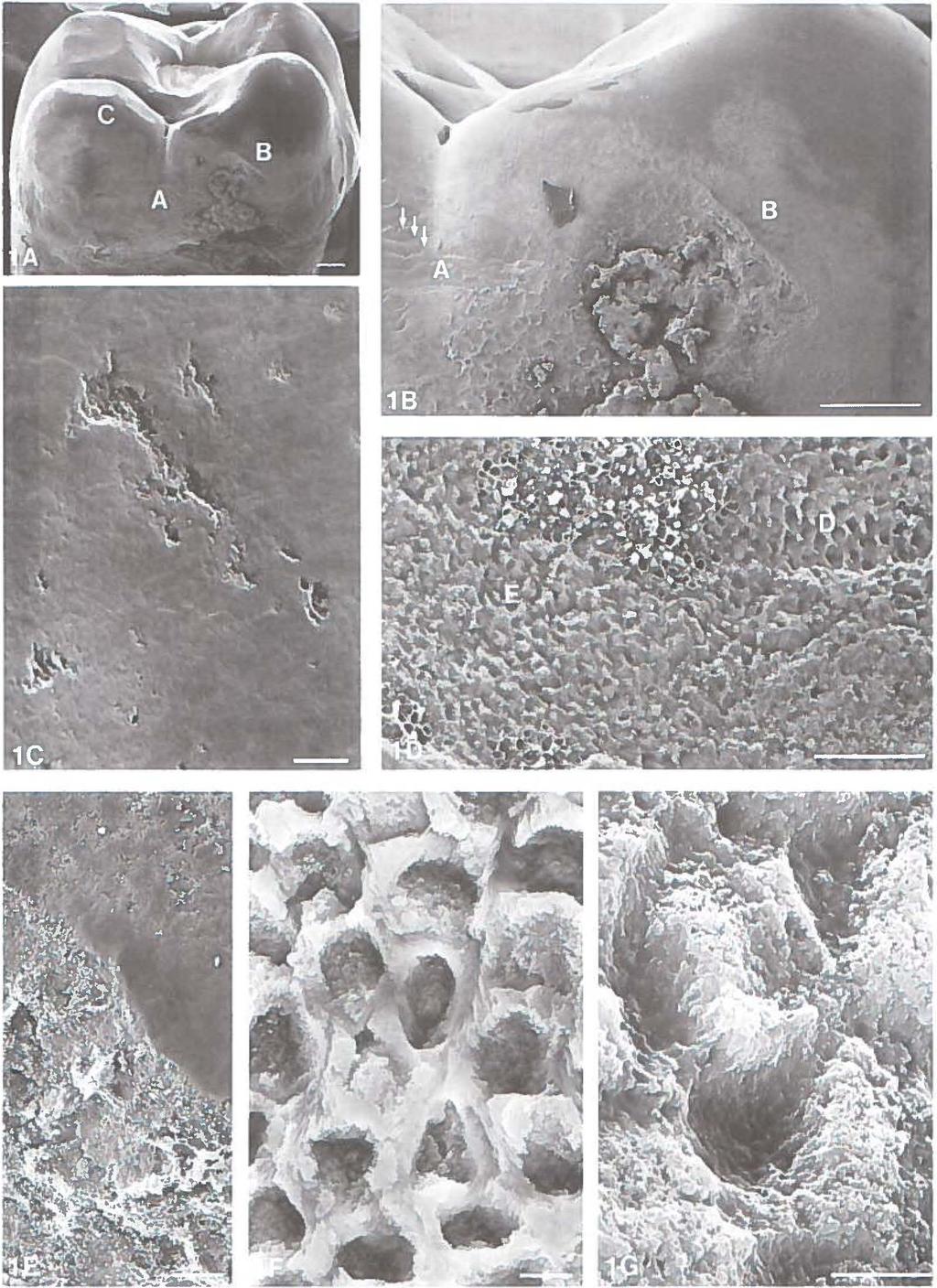
94 78
95 RESULTS All irradiated patients suffered from moderate to severe xerostomia with the amount of saliva in the oral cavities being less than 450 mg. The control subjects had amounts of saliva greater than 1.5 g in their oral cavity, which can be considered to be normal.19 Natural radiation caries Characteristic morphological features of natural radiation caries are depicted in SEM micrographs from a representative maxillary molar (Fig. 1), incisor (Fig. 2), and cuspid (Fig. 3). At most carious sites the teeth were covered with a dense plaque accumulation, consisting of spheroidal, spherical, rod-shaped and filamentous bacteria. Figure 1: maxillary molar. The distal surface of a maxillary molar is shown in Figs. la and lb. The neighboring tooth was removed before irradiation. The crown of the molar is relatively intact, but signs of superficial smooth surface decay can be seen on almost the entire distal surface (Fig. lb, areas A and B) and the cusps (Fig. la, area C). The pattern of decay is widespread and irregular. At location A demineralization resulted in the exposure of well pronounced perikymata (Fig. lb, arrows). Higher magnifications of this region (Figs. 10, lf, 1G) show a key hole like appearance with two patterns of destruction. The most characteristic type is due to preferential dissolution of the prism cores whereas interprismatic substance is less affected (area D on Fig. 10, Fig. lf). Similar hollowing out of prism cores can be observed at location B which is depicted at a higher magnification in Fig. le. Instead of hollowing out, a peripheral dissolution of prism sheaths with simultaneous dissolution of the interprismatic space was observed in some regions of the surface area (area E on Fig. 10, Fig. lg). A higher magnification of a functional cusp tip area (area C on Fig. la) shows porosity of enamel and crater formation (Figs. 1C). Subsurface enamel is exposed due to demineralization of the surface enamel at these locations. Figure 2: maxillary incisor. Fig. 2A shows a severely decayed maxillary incisor with extensive cervical caries. Most enamel is lost from the cervical and proximal areas. It is a characteristic finding that in the more advanced cases of radiation decay fractures occur within the enamel and large parts of the tooth crowns become Fig. I Maxillary molar. A Distal surface of a maxillary molar (Bar 1 mm). B Higher magnification of distal surface with smooth surface demineralization at location A with exposure of well pronounced perikymata (arrows) (Bar I mm). C Higher magnification of a functional cusp tip area at location C showing porosity of enamel and crater formation (Bar 50 Jlm). D Higher magnification of location A with key hole like appearance. Two different patterns of demineralization at locations D and E. (Bar 50 Jlm). E Higher magnification of location B showing porosity and crater formation on a smooth surface and hollowing out of prism cores in an area with large plaque deposits (Bar 50 Jlm). F Preferential dissolution of prism cores with remnants of interprismatic substance at location D (Bar 3 Jlm ). G Dissolution of prism sheaths as obsetved at location E (Bar 3 Jlm). 79
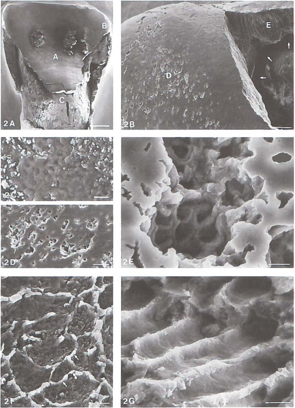
96 80
97 denuded of enamel resulting in exposure of the underlying dentin. The gap between enamel and dentin shown in the SEM micrographs (Fig. 2B, arrows) is an artefact caused by differences in shrinkage of enamel and dentin during preparation of the specimens for SEM. The palatal (area A on Fig. 2A, Fig. 2C) and incisal (area D on Fig. 2B, Fig. 20) surfaces show evidence of demineralization. The enamel in these regions shows large areas with irregular patterns of surface destruction with crater formation. Exposure of subsurface enamel in such craters with evidence of hollowing out of prism cores is depicted at a higher magnification in Fig. 2E. Fig. 2F represents an area of exposed dentin at the cervical region (area Con Fig. 2A). In most of these cases it looked as if the enamel mineral had dissolved from some areas and had reprecipitated in adjacent areas (Fig. 2F, arrows). A higher magnification of an area where fracture has occurred within the enamel (area Eon Fig. 2B) shows preferential dissolution and hollowing out of prism cores (Fig. 2G and IF). Figure 3: maxillary cuspid. Fig. 3A depicts a severely decayed maxillary cuspid with major parts of the labial, incisal, palatal and mesial surfaces denuded of enamel. Both exposure and decay of dentin can be observed. At location A, a large area of surface enamel has been lost (Fig. 3A). The result is exposure of subsurface enamel with pronounced perikymata and preferential dissolution of prism cores with remnants of interprismatic substance (Figs. 3B, 3E). Higher magnifications of the labial surface (area Bon Fig. 3A) show wide-spread crater formation (Figs. 3C, 30). Areas of fractured enamel (Fig. 3F) and exposed dentin (Fig. 3G) show the same phenomena as in Fig. 2, namely typical patterns of hollowing out of prism cores (Fig. 2G) and reprecipitation of dissolved enamel mineral (Fig. 2F). Induced radiation caries SEM of the enamel slabs from irradiated patients all showed characteristic patterns of demineralization and decay. The SEM micrographs from representative enamel slabs obtained from different patients are depicted in Figs. 4 and 5. Figure 4: early stages. During the first weeks most enamel slabs showed an accentuation of an arcade-formed prismatic structure (Figs. 4A, 4B, Fig. 40 area A). In many slabs microfractures developed along these prismatic arcades (Fig. 4C, Fig. 40 area B). From the third week onward most of the samples showed a porous enamel surface and starting crater formation (Figs. 4E, 4F). Although plaque accumulation was greater at the peripheral parts of the slabs, the carious destruction Fig. 2 Maxillary incisor. A Palatal surface of a severely decayed maxillary incisor with extensive cervical caries, loss of enamel from cervical and proximal areas, enamel fractures (location B) and exposure of dentin (Bar 1 mm). 8 Detail of the incisal edge showing demineralization (location D) and fractured enamel (location E); gap (arrows) due to shrinkage (Bar 100 J.Lm). C Detail of demineralization at location A (Bar 10 J.Lm). D Higher magnification of location D with an irregular pattern of porosity and crater formation (Bar 30 J.Lm ). E Detail of location A with crater formation and exposure of subsurface enamel (Bar 5 J.Lm). F Exposed dentin at location C with characteristic precipitation of calcium phosphate salts (arrows)(bar 10 J.Lm). G Higher magnification of fractured enamel at location E showing preferential dissolution of prisms (Bar 5 J.Lm). 81

98
99 was extensive in both the central and peripheral parts of the slabs. Figure 5: advanced stages. After three to six weeks most enamel slabs were severely damaged and demineralized. In many slabs large parts of the surface enamel were lost exposing the deeper enamel layers (area A on Fig. SA). At higher magnification a characteristic pattern of demineralization, namely preferential dissolution of the prism cores with remnants of interprismatic substance, was observed. The result is a key hole like appearance (Figs. SB, SC). Fig. SD represents a detail of area B on Fig. 5A, where generalized porosity with starting crater formation is depicted. In some cases the enamel had even peeled off exposing the dentin (Fig. 5E). The pictures suggest that the tooth mineral was dissolved from some areas and was reprecipitated in adjacent areas (Figs. SE, SF). Most control enamel slabs showed no demineralization during the twelve week experimental period when viewed in the SEM. At most, local areas with slight porosity of the enamel surface were observed. Hardness measurements. The changes in indentation length, I (J.Lm), ofthe enamel slabs placed in the oral cavities of irradiated patients (0-6 weeks) and control subjects (0-12 weeks) are shown in Fig. 6. In irradiated patients, about one third of the enamel slabs was damaged so severely by the progressive caries process after the third week that further measurements were not reliable. For that reason median values are presented instead of mean values, and no standard deviations are given. The hardness measurements indicated a significant demineralization of the samples in irradiated patients within 2 weeks. In control samples no increase in indentation length was observed. DISCUSSION Early radiation caries lesions seem to be similar to incipient normal caries lesions in permanent teeth The prismatic pattern of demineralization and the irregular pattern of destruction have also been observed in normal incipient carious lesions.21 Striking differences between normal and radiation caries are the rapid onset and progression of radiation caries, its widespread occurrence on enamel surfaces, the loss of large enamel parts and the fact that radiation caries is most commonly found on tooth surfaces that are relatively immune to dental caries. As mentioned in the introduction, the main etiological factor for radiation caries Fig. 3 Maxillary cuspid. A Severely decayed maxillary cuspid with major parts of labial, incisal, palatal and mesial surfaces denuded of enamel (Bar 1 mm). 8 Higher magnification of location A with loss of large part of surface enamel exposing subsurface enamel with pronounced perikymata (Bar 50 J.Lm). C Higher magnification of labial smooth surface at location B showing demineralization (Bar 100 J.Lm). D Higher magnification of location B with porosity of enamel and crater formation (Bar 10 J.Lm). E Detail of location A showing characteristic hollowing out of prism cores with remnants of interprismatic substance (Bar 10 J.Lm). F Higher magnification of fractured enamel at location D showing a longitudinal view with evidence of preferential prism dissolution (Bar 10 J.Lm). G Higher magnification of exposed dentin at location C with characteristic precipitation of calcium phosphate salts (arrows)(bar 10 J.Lm). 83
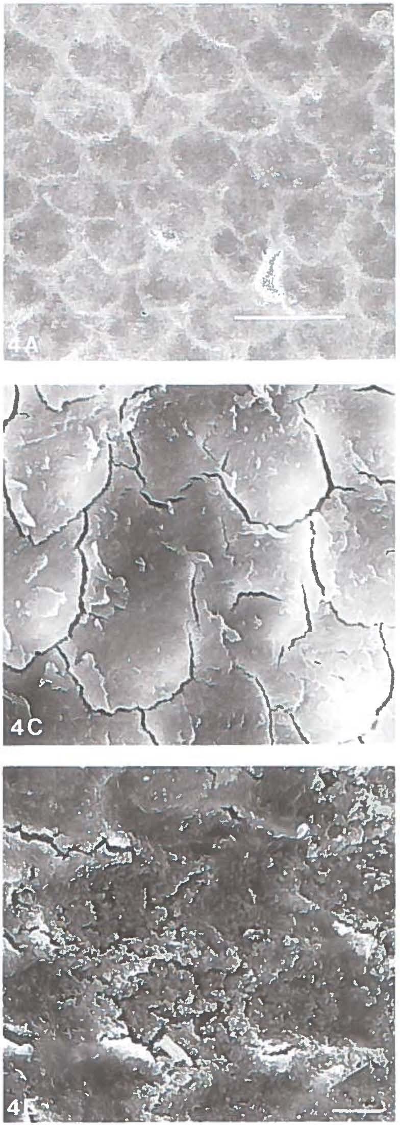

100 84
101 is irradiation-induced hyposalivation. The quantitative and qualitative changes of whole saliva and the concomitant shift in the oral flora towards cariogenic microorganisms produce a cariogenic environment that is aggravated by the changed pattern of food consumption.8 Direct effects of ionizing irradiation on dental enamel do not contribute significantly to the demineralization process In radiation caries the characteristic demineralization on caries immune, self-cleansing areas, is also related to the altered oral environment produced by irradiation-induced changes in salivary flow, composition and consistency of saliva that gives rise to accumulation of highly acidogenic dental plaque on these surfaces.3 The aggressiveness of the oral environment was clearly demonstrated in the model by the difference in progression between caries induced in irradiated versus non-irradiated subjects. To induce radiation caries lesions in a relatively standardized and reproducible way, the surface layer of the enamel slabs was removed in the model. Previous studies carried out in our laboratory had shown that the enamel fluoride concentrations in adjacent sites on unground surfaces varied significantly, whereas removal of the surface enamel by grinding resulted in enamel fluoride concentrations in adjacent sites that were not significantly different.24 The occurrence of dissolution of prism sheaths in some surface areas of extracted teeth in stead of the mostly observed hollowing out of prisms, may be related to local differences in the fluoride content of the surface layer25 and was probably seen less in the model because of removal of the surface layer during slab preparation. Edentulous subjects were chosen because data obtained in these subjects are not influenced by variables such as oral hygiene or other preventive measures (topical fluoride applications) which an irradiated dentulous subject has to apply in order to preserve his dentition. Considering the oral flora, only minor differences in its composition exist between dentulous and denture-wearing edentulous xerostomic patients.26 From the rapid, standardized and reproducible induction of radiation caries in the in situ model, the sequence of the stages in the development of radiation caries can be followed. The initial step in the demineralization process is the occurrence of porosity on widespread areas of enamel. This is followed by crater formation with exposure of subsurface enamel, preferential dissolution and hollowing out of prism cores, loss of large parts of surface enamel and loss of full enamel coverage exposing the underlying dentin. The same morphological features were observed in the extracted teeth with natural radiation caries. In these teeth, however, the more advanced stages predominated. In a previous investigation, comparison of results obtained with scanning longi- Fig. 4 Early stages of induced radiation caries. A Accentuation of arcade-formed prismatic structure (Bar 10 JLm). 8 Detail of accen!uated arcade-formed prismatic structure (Bar 3 JLm). C Development of splits and microfractures along prismatic arcades (Bar 3 JLm). D Starting crater formation from third week onward (Bar 10 JLm). E Higher magnification of crater formation at location B of Fig. 4D (Bar 3 JLm). F Higher magnification of craters from another slab showing exposure of deeper enamel layers (Bar 10 JLm). 85
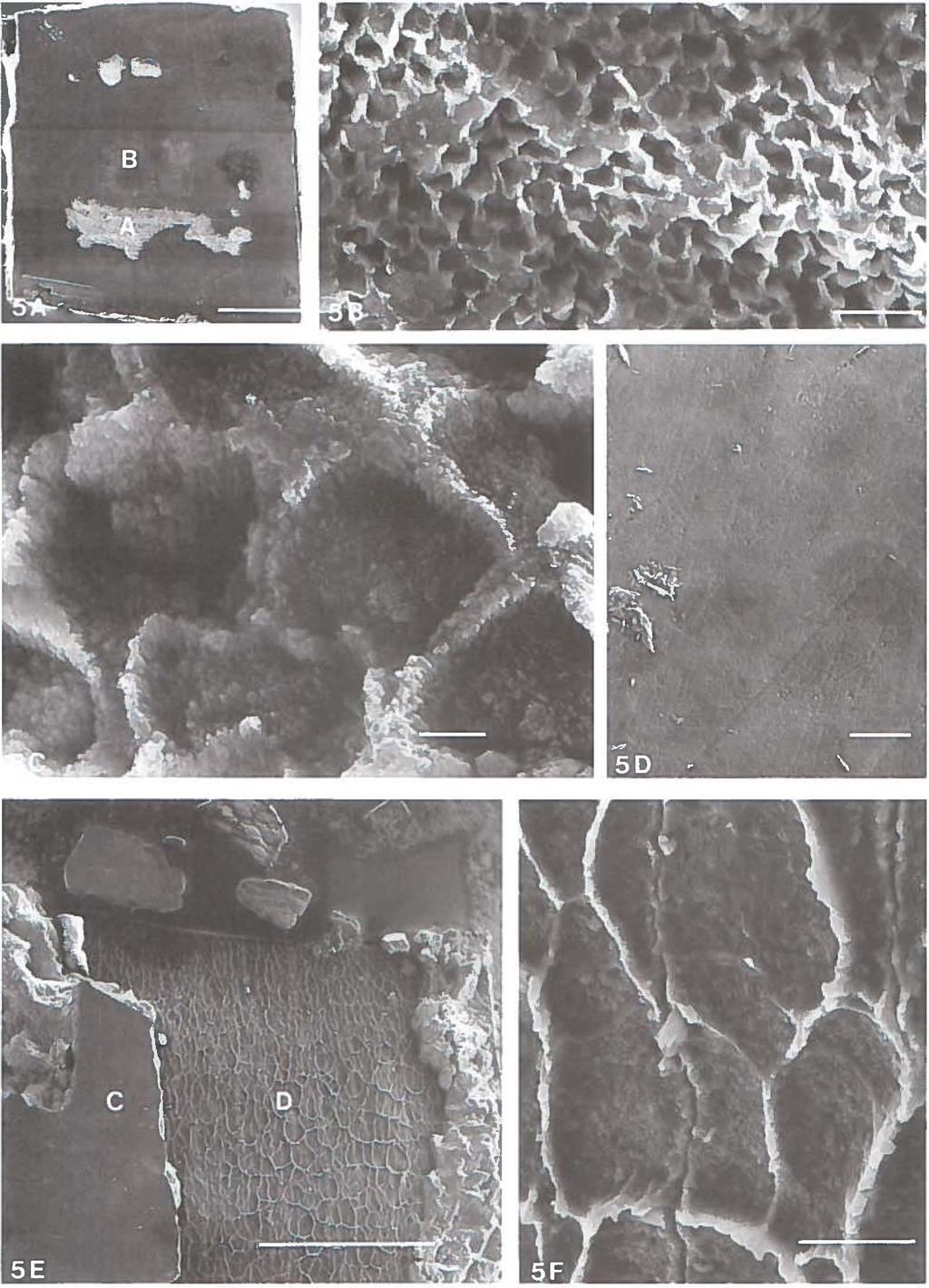
102 86
 of the enamel slabs in irradiated patients (n=7) and control subjects (n=6) as a function of time of demineralization (weeks).")
103 indentation length (1Jml _... irradiated subjects --controls t demin (weeks) Fig. 6 Indentation length (J.Lm) of the enamel slabs in irradiated patients (n=7) and control subjects (n=6) as a function of time of demineralization (weeks). tudinal microradiography (LMR), scanning optical monitoring (OM) and hardness measurements showed a significant increase in indentation length before any significant change in mineral composition could be observed with LMR and OM. 13 SEM observations only showed accentuation of prisms at that point of time. Although hardness measurements are not reliable on severely damaged enamel surfaces, they can be utilized as an indicator of the initial softening of the enamel slabs and were added to the model to characterize the onset of the caries process. In this study it has been shown that in situ induced radiation caries resembled natural radiation caries. In all patients, the induced radiation caries showed the same typical patterns of decay and the same sequence of events. The in situ model may offer an outstanding opportunity to study preventive regimens, for example fluoride applications, in preserving the natural dentition in subjects who have undergone head and neck radiotherapy. Fig. 5 Advanced stages of induced radiation caries. A Overview of enamel slab after three weeks with loss of large parts of surface enamel (Bar 1 mm). 8 Detail (area A) of subsurface enamel showing a characteristic prismatic pattern of demineralization with preferential dissolution of prisms and remnants of interprismatic substance (Bar 30 J.Lm). C Volcano aspect of hollowed out prism cores, detail of Fig. 58. (Bar 3 11-m). D Higher magnification of location B with generalized porosity and starting crater formation (Bar 3 J.Lm). E Overview of enamel slab after six weeks with evidence of loss of full enamel coverage at location D and relatively intact surface enamel at location C (Bar 1 mm). F Higher magnification of exposed dentin at location B with characteristic precipitation of calcium phosphate salts (Bar 30 J.Lm). 87
104 REFERENCES del Regato JA. Dental lesions observed after roentgen therapy in cancer of the buccal cavity, pharynx and larynx. Am J Roentgenol 1939;42: Frank RM, Herdly J, Philippe E. Acquired dental defects and salivary gland lesions after irradiation for carcinoma. J Am Dent Assoc 1965;70: Karmiol M, Walsh RF. Dental caries after radiotherapy of the oral regions. JAm Dent Assoc 1975;91: Ben-Aryeh H, Gutman D, Szargel R, Laufer D. Effects of irradiation on saliva in cancer patients. lnt J Oral Surg 1975;4: Brown LR, Dreizen S, Handler S, Johnston DA. Effect of radiation-induced xerostomia on human microflora. J Dent Res 1975;54: Brown LR, Dreizen S, Rider U, Johnston DA. The effect of radiation-induced xerostomia on saliva and serum lysozyme and immunoglobulin levels. Oral Surg Oral Med Oral Pathol 1976;41: Brown LR. Dreizen S, Daly TE. Drane JB, Handler S, Riggan U, Johnston DA. Interrelations of microorganisms, immunoglobulins and dental caries following radiotherapy. J Dent Res 1978;57: Dreizen S, Brown LR, Handler S, Levy BM. Radiation-induced xerostomia in cancer patients. Effects on saliva and serum electrolytes. Cancer 1976;39: Shannon IL, Starcke EN, Wescott WB. Effect of radiotherapy on whole saliva flow. J Dent Res 1977;56: Shannon IL, Wescott WB, Starcke EN, Mira J. Laboratory study of cobalt-60-irradiated human dental enamel. J Oral Med 1978;33: II Cowman RA, Baron SS, Glassman AH, Davis ME, Strosberg AM. Changes in protein composition of saliva from irradiation-induced "erostomia patients and its effect on growth of oral streptococci. J Dent Res 1983;62: Joyston-Bechal S. The effect of X-radiation on the susceptibility of enamel to an artificial caries-like attack in vitro. J Dent 1985;13: Jansma J, Buskes JAKM, Vissink A, Mehta DM, 's-gravenmade EJ. The effect of X-ray irradiation on the demineralization of bovine dental enamel. A constant composition study. Caries Res 1988;22: Markitziu A, Weshler R, Grajover M, Avital M, Gedalia A. Solubility, wear and microhardness in irradiated rat molars. J Dent Res 1989;68: Silverman S, Chierici A. Radiation therapy of oral carcinoma. Effects on oral tissues and management of the periodontium. J Periodontol 1965;36: Dreizen S, Brown LR, Daly E, Drane JB. Prevention of xerostomia related dental caries in irradiated cancer patients. J Dent Res 1977;56: Anneroth G, Holm LE, Karlsson G. The effect of radiation on teeth. A clinical, histological and microradiographic study. Int J Oral Surg 1985;14: Jongebloed WL, 's-gravenmade EJ, Retief DH. Radiation caries. A review and SEM study. Am J Dent 1988;4: Vissink A, 's-gravenmade, Panders AK, Vermey A, Petersen JK, Visch LL, Schaub RMH. A clinical comparison between commercially available mucin- and CMC-containing saliva substitutes. Int J Oral Surg 1983;12: Jansma J, Vissink A, 's-gravenmade EJ, de Josselin de Jong E, Jongebloed WL, RetiefDH. A model to investigate xerostomia-related dental caries. Caries Res 1988;22: Haikel J, Frank RM, Voegel JC. Scanning electron microscopy of the human enamel surface layer of incipient carious lesions. Caries Res 1983;17: Holmen L, Thylstrup A, Ogaard B, Kragh F. A scanning electron microscopic study of progressive stages of enamel caries in vivo. Caries Res 1985;19: Jansma J, Borggreven JMPM, Driessens FCM, 's-gravenmade EJ. Effect of X-ray irradiation on the permeability of bovine dental enamel. Caries Res 1990;24: Benediktsson S, Retief DH, Bradley EL, Switzer P. The effect of contact time of acidulated phosphate fluoride on fluoride concentration in human enamel. Archs Oral Biol 1982;27: Jongebloed WL. An ultrastructural study of the caries process; thesis Groningen, Weerkamp AH, Wagner K, Vissink A, 's-gravenmade EJ. Effect of the application of a mucin-based saliva substitute on the oral microflora ofxerostomic patients. J Oral Pathol l987;16:
105 IN SITU STUDY ON THE PREVENTION OF RADIATION CARIES INTRODUCTION Radiation caries, a rapidly progressing highly destructive type of dental caries, is a common side effect of radiation treatment.1 3 According to Dreizen et al,4 carious lesions can be seen within three months and extreme damage of the dentition within one year after the start of radiotherapy. In several clinical studies it has been shown that almost complete caries prevention can be achieved in irradiated patients by the daily application of topical fluoride (F) agents combined with a program of strict oral hygiene.4 9 The aim of this study was to develop an optimal preventive program for radiation caries by evaluating the effects of F concentration and application procedures in subjects who have had irradiation treatment for malignant tumors of the head and neck. MATERIALS AND METHODS Subjects. Seven edentulous subjects (3 women and 4 men) suffering from radiationrelated xerostomia participated in this study. The subjects received an average radiation dose of 55 Gy (range Gy) at a level of 2 Gy/day, 5 days/week from a 60Co source. The mean age of the subjects was 67.3 years (range yrs). All subjects wore full dentures. The severity of xerostomia was measured by wiping the oral cavity after swallowing with a water-absorbent gauze, which was weighed before and after saliva collection. The tests were performed on three different days between and a.m., and the subjects were not allowed to take food or beverages for 2 h before the test.10 Experimental design. In this study a recently developed in situ model for the investigation of xerostomia-related dental caries was used.11 Both the left and right molars of the lower denture of each subject were replaced by a metal sample holder.12 Each holder contained six human enamel slabs. The slabs could be removed and replaced by unscrewing the occlusal part of the holder (Fig. 1 ). About 9 mm2 of each slab was exposed to the oral environment. In all subjects four experiments were performed: procedure A: no F therapy (control); procedure B: 1% neutral NaF gel applied for 5 min every 2nd day; procedure C: 1% neutral NaF gel applied for 5 min once a week, and procedure D: rinsing with 10 ml of a F containing mouthwash (0.05% NaF, Prodent, Amersfoort, The Netherlands) for 1 min once daily. The subjects received information sheets with instructions for each of the four procedures. The NaF gel was applied with a squeeze 89

106 Fig. 1 Sample holder mounted in a lower denture. The slabs can be removed and replaced by unscrewing the occlusal part of the holder. bottle (one drop per enamel specimen). The subjects were not allowed to clean the enamel slabs. They were instructed to keep their dentures in tap water during the night. The enamel slabs were analyzed at weekly intervals using longitudinal microradiography (LMR), scanning optical monitoring (OM), scanning electron microscopy (SEM) and hardness measurements (HM). F analyses of the enamel slabs were done prior to their insertion in the appliances and after 6 weeks intraoral exposure. Each experiment extended over 6 weeks and the next was started after an interval of 2 weeks. In each experiment 12 enamel slabs per subject were examined: 4 for LMR and OM, 6 for HM and SEM and 2 for F analyses. Slabs used for SEM were replaced by acrylic blocks. Enamel slab preparation. The facial enamel surfaces of noncarious human mandibular permanent incisors were partially ground flat on 1,200 grit silicon carbide paper, polished on a Kent Mark II polisher (Engis, Maidstone, England) using Hypress diamond compounds (Engis) and cut in rectangular slabs (3 x 4 x 1.5 mm) by means of a water-cooled diamond saw (Horico, Berlin, FRG). For LMR and OM the lingual aspects of the enamel slabs were ground on 220 grit silicon carbide paper to obtain planoparallel slabs with a thickness of 340 ± 20 IJ.m. All enamel slabs were embedded in cold-cure polymethylmethacrylate (de Trey, Wiesbaden, FRG) and ultrasonically cleaned in tap water for 10 min. Care was taken to keep the experimental facial sides free from acrylic resin. LMR and scanning OM. By means of dental impression paste (President Regular Body; Coltene, Altstiitten, Switzerland) the enamel slabs for LMR and OM were 90

107 embedded in polymethylmethacrylate sample holders which fitted in both the LMR and OM experimental setup. In this manner an enamel slab could exactly be repositioned in its individual holder at each measuring interval to be scanned with LMR and OM at the same discrete surface position. LMR was performed as described by de Josselin de Jong13 and de Josselin de Jong et al For OM the optical caries monitor as described by ten Bosch et al and Borsboom and ten Bosch18 was applied. Both methods were performed before the start of each experiment and at weekly intervals for the duration of the investigation. CROSS SECTION A c Fig. 2 Apparatus for the measurement of F. A Selective F electrode, 8 reference calomel electrode, C suction apparatus. Scanning electron microscopy. The enamel slabs were washed in running tap water to remove surface debris, and glued on aluminum stubs with fast-curing epoxy resin. When transverse examinations were required, these enamel slabs were also fractured. A thin Au layer (approximately 15 nm) was sputtered on the slabs. Scanning electron micrographs were taken at weekly intervals with a JEOL Type 35C (JEOL, Tokyo, Japan) scanning electron microscope operated at 25 kv. Hardness measurements. Microhardness measurements were performed with a Leitz Durimet miniload hardness tester with a Knoop diamond (Leitz, Wetzlar, FRG) at a load of 100 g, applied for 20 s. Five indentations were made in a definite 91
108 Jmlkg.m , t lwkl Fig. 3 Mineral loss, (kg.m-2 ), of the enamel samples as a function of the time of demineralization (weeks) in the four experiments. Median values (n=7). = No F therapy; = F gel 1/2 days; e = F gel l/week; = F mouthrinse 1/day. pattern in the central area of each enamel slab. The measurements were performed at weekly intervals. Biopsy procedures and F analysis. Three successive acid etch biopsies were performed on the ground enamel surface of each enamel slab prior to insertion in the intraoral device. Biopsy sites were demarcated by placing an adhesive tape with a circular hole of 1 mm diameter on the enamel surface. Then 0.4 J.LI of 1 M perchloric acid was deposited on the demarcated biopsy site, and absorbed after 5 s with a filter paper disc which was placed in a polyethylene tube containing 25 J.Ll total ionic strength adjustment buffer (TISAB) (Orion Research, Cambridge, Mass., USA). The etched area was washed twice in quick succession with 0.4 J.Ll TISAB and the washings transferred to the polyethylene tube. The F concentrations in 5-J.LI volumes of the etching solutions were determined by a microanalytical technique developed by Vogel et al. 19 The apparatus consists of a F selective electrode (Orion Research; Fig. 2,A) and a calomel reference electrode (Fig. 2,B) linked by a microcapillary tube. The phosphorus concentrations were determined in 10 J.LI volumes by the analytical technique developed by Chen et al.20 using a Spectronic 2000 spectrophotometer (Bausch and Lomb, Rochester, N.Y., USA). The mass enamel in the etching solutions was calculated by assuming that enamel contains 18.0% P,21 and expressed in micrograms. The enamel F concentrations were adjusted to standardized depths of 5 J.Lm.22 After the enamel slabs were exposed to the intraoral environment for 6 weeks, three successive acid etch biopsies were again carried out on demarcated biopsy sites immediately adjacent to the initial biopsy sites, and the enamel F concentrations were again adjusted to standardized depths of 5 J.Lm. Statistical analysis. As a result of the deterioration of the enamel blocks in the oral environment of the xerostomic patients, exact measurements for LMR, OM and 92
, as a function of the time of demineralization (weeks) in the four experiments. Median values (n=7).")
109 / --.-L: / o.lc!!'=,r :::l ; t!wkl Fig. 4 Change in optical scattering, "KS' ( mm -I ), as a function of the time of demineralization (weeks) in the four experiments. Median values (n=7). = No F therapy; t = F gel l/2 days; e = F gel l/week;.l = F mouthrinse 1/day. HM were sometimes not possible. These measurements were not omitted, but were considered as being extreme in an untoward direction. The median was used as the summary statistic for measurements from a single subject in a given week. An overall comparison of the results obtained after 6 weeks was accomplished by the generalized signed-rank test.23 When the results were significant at the 5% level, this test was followed by a pairwise signed-rank test (one-tailed at the 1% level). At a one-tailed 5% level, the latter was used to compare per experiment the results after 6 weeks to those prior to the exposure of the enamel blocks to the oral environment. Logarithms of the adjusted enamel F concentrations were used in the statistical analysis. For each enamel block and for each etch depth (0-5, 5-10, /Lm) the F acquired by the enamel was calculated by subtracting the adjusted baseline enamel F concentration (week 0) from the adjusted experimental enamel F concentration (week 6). The data were analyzed by means of a multivariate analysis of variance using the SYSTAT statistical package.24 RESULTS The subjects participating in this study suffered from moderate to severe xerostomia with the mean ± SD of the amount of saliva in the oral cavities being 414 ± 218 mg. Healthy subjects who do not use drugs and who have not been exposed to radiation therapy have 1,800-3,000 mg saliva in their oral cavities.10 Plaque accumulation on the enamel slabs was observed within 1 day. The composition of the oral flora was comparable to the flora in postradiation subjects with natural teeth.25 The mineral loss in Llill (kg.m-2) of the slabs exposed to the control and experimental disciplines is shown in figure 3. The changes in optical scattering KS (mm -I) are depicted in figure 4, and the results of the micro hardness tests are presented in figure 5. The ranges of variations of the median obtained with 93
110 l! ml 200 Fig. 5 Indentation length (1, JLm) as a function of the time of demineralization (weeks) in the four experiments. Median values (n=7). = No F therapy; + = F gel l/2 days; e = F gel 1/week;.t. = F mouthrinse 1/day. the LMR, OM and HM are shown in figure 6. In the control experiment (procedure A), significant demineralization of the slabs was measured with LMR, OM and HM. Application of F for 6 weeks (procedures B-D) resulted in significant inhibition of the demineralization. Evaluation by LMR and OM showed that the application of F gel every second day was significantly more effective in reducing the demineralization process than the other F therapies (Table 1}. Hardness measurements (HM) showed no significant differences among the three F therapies (Table 1 ). Table 1 Treatment differences (p values; one-tailed signed-rank test) Evaluation F-gel 1/2 days vs. F-gel 1/2 days vs. method F-gel 1/week F-mouthrinse 1/day F-gel 1/week vs. F-mouthrinse 1/day LMR OM HM >0.20 >0.20 >0.20 Great variations in the surface morphology of the enamel slabs were observed by SEM. After 3 weeks porous enamel surfaces, starting crater formation and hollowing out of prism cores were observed in most of the control enamel slabs after exposure to the oral environment. The caries process proceeded progressively resulting in severely demineralized slabs after 6 weeks (Fig. 7). In some of the slabs loss of enamel resulted in the exposure of dentin. Enamel slabs exposed to the F therapies showed a reduction in enamel demineralization when compared with the control slabs, but no clear differences were seen between the applied F therapies. In most 94
. Am (kg.")
111 .lni!kg.m 'l LMR :IS!mm l l 1 l OM.... Xi l ml HM 0 A o o- 0 0 AA O.B a A 0 A- "' A o- - A A o- 0 A- 0 & A A 00 0 A A A Fig. 6 Range of median values as obtained with LMR, OM and HM at week 6 (n=7). Am (kg.m-2 ), (mm - 1 ) and LTI (I'm) indicate the change in, respectively, mineral content, optical scattering and indentation length. 0,. = No F therapy; 0 = F gel 1/2 days; O,e = F gel 1/week; 6,11. = F mouthrinse 1/day. Arrows point to the median values as shown in figure 3-5;. e and 11. = severely damaged enamel slabs. slabs only the initial stage of enamel demineralization was observed (Fig. 8). The median adjusted F concentration of the enamel slabs at the three depths studied prior to the insertion in the intraoral device (week 0) is given in Table 2. The adjusted baseline enamel F concentrations at the three etch depths were not significantly different (p>o.lo). The adjusted experimental enamel F concentrations increased in the 44 enamel slabs exposed to the control and F therapies for 6 weeks (p<o.ol). Enamel demineralization was so severe in 5 of the enamel slabs that acid etch biopsies could not be performed. This increase varied significantly among subjects (p<o.ool), among the various procedures (p<o.ool), and at different depths (p<0.05), but the interactions between the three parameters, subjects, procedures and etch depths were not significant (p>o.lo). The increase of enamel F concentration at the first etch depth was estimated to be 1.2 times greater than the increase at the third etch depth. The increase in enamel F concentration depends only slightly on etch depths and can conveniently be described by one number. The 'median increase per enamel slab' observed in the different treatment procedures is presented in Table 3. In the control slabs the enamel F concentration increased by a factor of 1.5; on exposure to the F therapies the factors ranged from 3.1 to 4.1. The differences between the control and F procedures were significant (p<o.ool ). The factor obtained for the application of F gel every 2nd day was significantly different from the factor for the weekly application of the F gel (p<0.05). 95
. Fig. 8 F-treated enamel slab after 6 weeks.")
.")
112 Fig. 7 Overall view of an untreated enamel slab after 6 weeks. Presence of a 'relatively' unaffected enamel layer at location A, which seems to be loosening of the deeper part of the slab. Complete absence of enamel and the exposure of the underlying dentin at location B. The cracks in the slabs are artefacts (Bar 1 mm). Fig. 8 F-treated enamel slab after 6 weeks. Porosity of enamel surface and starting crater formation (double arrow) can be observed. Single arrows mark the border line of the exposed part of the slab. Cracks are artefacts in the sample (Bar 1 mm). DISCUSSION Previous studies carried out in our laboratory showed that the enamel F concentrations in adjacent sites on unground surfaces varied significantly.26 Removal of the surface enamel by grinding resulted in the enamel F concentrations in adjacent sites being not significantly different. This was the reason that ground enamel slabs were used in the present study. Intradental control was therefore possible and the 96
113 Thble 2 Median F concentration (ppm F) of the enamel slabs at week 0 (n = 49) Etch depth Minimum value Percentile Maximum value 25th 50th 75th p.m Ta ble 3 Median F uptake per enamel block, (F- 6w ]I(F-0w ], under the different experimental conditions Experiment No F F-gel 112 days F-gel 1/week F-mouthrinse (F-6w]I(F- Ow] 1.5* 4.1** % confidence limits p<0.001: (no F vs. F experiments); p<0.05: (F-gel 1/2 days vs. F-gel l/week). F acquired at each etch depth was obtained by subtracting the adjusted baseline enamel F concentrations (0 weeks) from the corresponding adjusted experimental F concentrations (6 weeks). It is apparent from the results that application of F resulted in significant reduction of demineralization. With both LMR and OM it was shown that F gel applied every second day was significantly more effective than the other F therapies. The differences in the LMR and OM values (positive versus negative values; F gel 1/2 days, Figs. 3, 4 and 6) may be interpreted as slight depositions of organic material on the surface. This translucent layer did not influence the LMR data, while it diminished optical scattering S. F analysis demonstrated a significantly higher F uptake after F gel applied every 2nd day compared to the weekly gel therapy. Hardness measurements, however, did not show significant differences between all therapies, although there was a tendency which indicated a greater inhibitory effect of F gel applied every 2nd day. This may be explained by the fact that indentation length on whole samples is mainly a qualitative parameter for the outer enamel region. In case of subsurface lesions, indentation length does not give details of the hardness changes below the surface nor in different regions of the lesion.27 Although F gel applied every 2nd day was the most effective therapy in this study, slight demineralization of enamel was still observed. The necessity for additional oral hygiene measures is stressed by this observation. This is in accordance with reported data that complete radiation caries prevention can be accomplished with the use of high-dose F therapies and strict adherence to oral hygiene procedures F mouthrinses used once daily, which are easy to per- 97
114 form, could result in a much higher degree of patient compliance.6 In the present study the daily use of fluoride mouthrinses was only effective in the subjects with moderate demineralization of the enamel slabs. In all other subjects the use of F mouthrinses once daily was inadequate to prevent demineralization. It is, however, difficult to define the caries susceptibility at the onset of the preventive treatment. Therefore, the application of a F gel every 2nd day and a strict oral hygiene regimen are recommended as an optimal procedure to inhibit the onset of radiation caries. From this study it may be concluded that F gel applied every 2nd day was the method of choice among those tested for preventing the onset of radiation caries. REFERENCES del Regato JA. Dental lesions observed after roentgen therapy in cancer of the buccal cavity, pharynx and larynx. Am J Roentgenol 1939;42: Frank RM, Herdly J, Philippe E. Acquired dental defects and salivary gland lesions after irradiation for carcinoma. JAm Dent Assoc 1965;70: Karmiol M, Walsh RF. Dental caries after radiotherapy of the oral regions. JAm Dent Assoc I975;9I: Dreizen S, Brown LR, Thomas TE, Drane JB. Prevention of xerostomia-related dental caries in irradiated cancer patients. J Dent Res I977;56: Wescott WM, Starcke EN, Shannon IL. Chemical protection against postirradiation dental caries. Oral Surg Oral Med Oral Pathoi i975;40: Katz S. The use of fluoride and chlorhexidine for the prevention of radiation caries. J Am Dent Assoc I982;I04:I Markitziu A, Gedalia I, Stabholz A, Shuval J. Prevention of caries progress in xerostomic patients by topical fluoride applications: a study in vivo and in vitro. J Dent 1982;10: Horiot JC, Schraub S, Bone MC, Bain Y, Ramadier J, Chaplain G, Nabid N, Thevenot B, Bransfield D. Dental preservation in patients irradiated for head and neck tumours: A IO-year experience with topical fluoride and a randomized trial between two fluoridation methods. Radiother Oncol I983; 1: Meyerowitz C, Featherstone JDB, Shariati M, Eisenberg AD. Radiation-induced hyposalivation and caries. J Dent Res I986;65:831. IO Vissink A, 's-gravenmade EJ, Panders AK, Vermey A, Petersen JK, Visch LL, Schaub RMH. A clinical comparison between commercially available mucin- and CMC-containing saliva substitutes. Int J Oral Surg 1983;12:132-I38. II Jansma J, Vissink A, 's-gravenmade EJ, de Josselin de Jong E, Jongebloed WL, Retief DH. A model to investigate xerostomia-related dental caries. Caries Res I988;22: de Bruyn H, Hummel M, Arends J. In vivo effect of a fluoridating varnish with various fluoride contents on human enamel. Caries Res I985;19: de Josselin de Jong E. Comparison of methods in caries research; thesis Groningen, I986. I4 de Josselin de Jong E, van der Linden AHIM, Borsboom PCF, ten Bosch JJ. Determination of mineral changes in human dental enamel by longitudinal microradiography and scanning optical monitoring and their correlation with chemical analysis. Caries Res I988;22:I de Josselin de Jong E, van der Linden AHIM, ten Bosch JJ. Longitudinal microradiography: A new nondestructive quantitative method to follow mineral changes in mineralized tissue slices. Phys Med Biol 1987;32: ten Bosch JJ, Borsboom PCF, ten Cate JM. A nondestructive method for monitoring de- and remineralization of enamel. Caries Res 1980; 14: ten Bosch JJ, van der Mei HC, Borsboom PCF. Optical monitor of in vitro caries. A comparison with chemical and microradiographical determination of mineral loss in early caries lesions. Caries Res 1984;18: I8 Borsboom PCF, ten Bosch JJ. Fiber-optic scattering monitor for use with bulk opaque material. Appl Opt 1982;21:
115 19 Vogel GL, Chow LC, Brown WE. A microanalytical procedure for the determination of calcium, phosphate and fluoride in enamel biopsy samples. Caries Res 1983;17: Chen PS, Toribara TY, Warner H. Microdetermination of phosphorus. Anal Chern 1956;28: Siiremark R, Samsahl K. Gamma-ray spectrometric analysis of elements in normal human enamel. Arch Oral Bioi 1961;6: Retief DH, Sorvas P, Bradley E, Taylor R, Walker A. In vitro fluoride uptake, distribution and retention by human enamel after 1- and 24-hour application of various topical fluoride agents. J Dent Res 1980;59: Fidler V, Nagelkerke NJD. A generalized signed-rank test for comparison of p treatments. Statistica Neerlandica 1986; 40: Wilkinson L. SYSTAT: The system for statistics. (SYSTAT Inc., Illinois 1986). 25 Weerkamp AH, Wagner K, Vissink A, 's-gravenmade EJ. Effect of the application of a mucin-based saliva substitute on the oral microflora of xerostomic patients. J Oral Patho1 1987;16: Benediktsson S, Retief DH, Bradley EL, Switzer P. The effect of contact time of acidulated phosphate fluoride on fluoride concentration in human enamel. Archs Oral Bioi 1982;27: Featherstone JOB, ten Cate JM, Shariati M, Arends J. Comparison of artificial caries-like lesions by quantitative microradiography and microhardness profiles. Caries Res 1983;17: Reynolds WR, Hickey A, Feldman MI. Dental management of the cancer patient receiving radiation therapy. Clin Prev Dent 1980;2:
116
117 Chapter 4 A SURVEY OF THE PREVENTION AND TREATMENT REGIMENS OF ORAL SEQUELAE RESULTING FROM HEAD AND NECK RADIOTHERAPY USED IN DUTCH RADIOTHERAPY INSTITUTES This chapter is based on the following publication: Jansma J, Vissink A, Bouma J, Vermey A, Panders AK, 's-gravenmade EJ. A survey of the prevention and treatment regimens of oral sequelae resulting from head and neck radiotherapy used in Dutch radiotherapy institutes. lnt J Radial Oneal Bioi Phys (submitted, 1991). 101
118 INTRODUCTION The incidence of malignant head and neck tumors (skin cancer excluded) in the Netherlands is about 23/100,000 inhabitants, i.e. circa 3500 new cases per year.1 Radiation treatment plays an important role in the management of head and neck cancer. According to Rothwell2 approximately 50% of all new cases of invasive head and neck cancer will need radiotherapy as a primary treatment, as an adjunct to surgery or chemotherapy or as palliation. The radiation dose needed for curative treatment is based on the location and the type of malignancy and whether or not radiotherapy will be used alone or as a pre- or post-surgical aid. Most patients who suffer from head and neck carcinomas receive between 50 and 70 Gy as a curative dose, which is usually given over a 5-7 week period, once a day, five days a week, with a daily tumor dose of about 2 Gy. Due to the location of the primary tumor and the regional lymph nodes, the oral cavity, salivary glands and jaws are in the field of radiation in most head and neck cancer patients, resulting in unwanted irradiation-induced changes in these tissues. The major side effects that may occur during or after radiotherapy in the head and neck region are mucositis, hyposalivation, taste loss, radiation caries and osteoradionecrosis ( ORN). These side effects may form a heavy burden for the patient, cause a lot of distress and have a tremendous impact on the quality of life. Mucositis is a transient complication, but it is an integral part of the radiation therapy in terms of morbidity. Mucositis causes local discomfort and pain as well as difficulties in drinking, eating, swallowing and speech during a period of about seven weeks. As a consequence nutritional problems can arise causing a bad nutritional condition and in severe cases nasogastric tube feeding may become necessary, which is very uncomfortable.3 4 Severe mucositis may even necessitate a break in the course of radiation treatment and can thus become a dose-limiting factor, with all its risks.2 5 Hyposalivation leads to distressing, longlasting, often irreversible complaints, such as oral dryness, hampered oral functioning, nocturnal oral discomfort, burning mouth, impeded social activities, reduced taste and high susceptibility to oral infections and dental caries.6 7 The effects of irradiation on the jaws lead to a lifelong risk for the development of ORN Fortunately, it is generally accepted nowadays that most oral complications of head and neck irradiation can be prevented or reduced in severity.11 Prevention regimens are, however, mainly based on clinical experience. The result is a great diversity in procedures, which may lead to a variety in the preventive approach in daily practice. No studies on the existence of such variety in our, or any other country have been published to date. The purpose of the present investigation was to survey the prevention and treatment regimens applied in all radiotherapy institutes in the Netherlands, and to evaluate the differences in these regimens. For practical reasons, this survey was not intended to evaluate the effects of the various regimens in the patient situ- 102
119 ation. Therefore no value judgements about specific care programs can be given other than by comparison with data from the literature. MATERIALS AND METHODS Dutch radiotherapy institutes. All centers in which irradiation for head and neck cancer is performed (n=20) were included in the study. They were all willing to cooperate and to make the study a success. According to the patient numbers reported by these centers, a total of about 2200 patients were treated with radiotherapy to the head and neck region in In this number, patients treated for laryngeal carcinoma and patients treated for (non) Hodgkin lymphoma in the head and neck region are included, patients with skin cancer are excluded. The participating institutes are listed in Table 1. Thble I Dutch radiotherapy institutes in which head and neck radiotherapy is performed Institute Academisch Medisch Centrum, Amsterdam Academisch Ziekenhuis Rotterdam Dijkzigt, Rotterdam Academisch Ziekenhuis Groningen, Groningen Academisch Ziekenhuis Leiden, Leiden Academisch Ziekenhuis Nijmegen St Radboud, Nijmegen Academisch Ziekenhuis Utrecht, Utrecht. Academisch Ziekenhuis Vrije Universiteit, Amsterdam Antoni Van Leeuwenhoek Ziekenhuis, Amsterdam Arnhems Radiotherapeutisch Instituut, Arnhem Catharina Ziekenhuis, Eindhoven Dr Bernard Verbeeten Instituut, Til burg Dr Daniel den Hoed Kliniek, Rotterdam Medisch Spectrum Twente, Enschede Radiotherapeutisch Instituut Friesland, Leeuwarden Radiotherapeutisch Instituut Limburg, Heerlen Radiotherapeutisch Instituut Stedendriehoek en Omstreken, Deventer Rode Kruis Ziekenhuis, 's-gravenhage St Sophia Ziekenhuis, Zwolle Westeinde Ziekenhuis, 's-gravenhage Ziekenhuis Leyenburg, 's-gravenhage Including patients of Radiotherapeutisch Instituut Zeeland, Vlissingen These institutes use the same radiotherapeutic unit, but were evaluated separately because of different preventive programs Survey. All centers were isited in the period June-October 1990 and at least two members of the staff responsible for the prevention and treatment of the oral side effects of head and neck irradiation were interviewed, i.e. radiotherapist and/or dental team (oral and maxillofacial surgeon, hospital dentist, oral hygienist). In centers without a dental team or dentist, only the radiotherapist was interviewed. 103
120 Table 2 Composition of dental teams in 20 radiotherapy institutes. In several centers more than one oral and maxillofacial surgeon, hospital dentist and/or oral hygienist were part of the team Oral and maxillofacial surgeon, hospital dentist and oral hygienist Oral and maxillofacial surgeon and oral hygienist Oral and maxillofacial surgeon Hospital dentist and oral hygienist No dental team 5/20* 8/20 1/20 3/20 3/20 5 out of 20 centers etc. Table 3 Percentage of patients in 20 radiotherapy institutes, in whom the oral cavity and/or the salivary glands were included in the field of radiation, referred to a dental team prior to radiotherapy Dentulous patients Edentulous patients All patients (100%) % of the patients 50-75% of the patients < 50% of the patients Only family dentist No screening for foci 9/20 4/20 3/20 1/20 3/20 0/20 7/20 4/20 2/20 4/20 0/20 3/20 All interviews were performed by the same interviewer (J.J.) using a list of open questions. The answers were scored by the interviewer according to predefined response categories. New categories were added when the response did not match the predefined ones (see appendix). In the result section 'n' always refers to the number of centers in which the respondents positively answered a certain item. Questions referred to: a composition of the team responsible for the prevention and treatment of radiation-related oral side effects b screening and care pre-irradiation: - moment of first contact of the patient with the team - dental assessment and screening on foci of infection extraction protocol instructions for oral hygiene and fluoride application c care during irradiation: - oral hygiene and mucositis protocol - fluoride protocol - denture wearing d care post-irradiation: - oral hygiene and fluoride protocol - denture wearing - extraction protocol treatment of hyposalivation - follow-up 104
121 RESULTS Composition of the dental team. In the majority of centers at least one oral and maxillofacial surgeon ( n = 14) or one oral hygienist ( n = 16) were part of the team, occasionally supplemented by a hospital dentist (n=8)(table 2). A team consisting of an oral and maxillofacial surgeon, an oral hygienist and a hospital dentist was only present in five centers. In three centers the pre-irradiation screening and preand post-irradiation care of the patients was left to their family dentist because of the absence of a dental team. In all centers a radiotherapist screened the patients on the development of oral side effects, particularly mucositis, at least once a week during radiation treatment. Screening and care pre-irradiation. A considerable number of patients in whom the oral cavity and/or salivary glands were included in the field of radiation, was not screened prior to radiotherapy by a dental team (Table 3). The centers in which no dental team was involved in counseling of at least 50% of the edentulous (n=7) and dentulous (n=4) patients were not the centers with the lowest patient numbers (569 patients in total, range patients). If patients were screened, they were always referred to the dental team prior to the onset of radiotherapy, and about fifty percent of them was screened more than two weeks before irradiation. The standard pre-irradiation dental assessments and instructions are presented in Table 4. In centers with a dental team ( n = 17), a panoramic X-ray was always made for screening on foci of infection in dentulous patients. Two of these centers made no X-rays in edentulous patients. No information was obtained in case patients were referred to their family dentist. Baseline data on composition of oral flora (n=o) and mouth opening (n=o) were not collected. In two centers the salivary flow rate was measured. Professional tooth cleansing was performed only in centers with an oral hygienist. Periodontal disease was occasionally treated with rootplaning and curettage in stead of extraction (n= 11), particularly when an oral hygienist was a member of the team (n=10). If extraction or surgical removal of teeth or root tips was indicated, a minimum interval of 0-1, 1-2 or 2-4 weeks between extraction and the onset of radiotherapy was considered necessary in one, nine and ten centers, respectively. Wound healing was routinely checked in 12 centers before irradiation was started. In the majority of the centers dentulous patients were instructed on toothbrushing (n= 16) and interdental cleansing (n=15). In edentulous patients oral hygiene and denture hygiene were instructed only in ten and twelve centers, respectively. Standard fluoride usage, other than by brushing with fluoridated toothpaste, was instructed in 18 centers. In two of them fluoride was prescribed by the radiotherapist. The fluoride preparations used are listed in Table 5. Self-application of the fluoride gel was prescribed in 11 centers, while in another four centers the gel was applied by an oral hygienist. Custommade carriers (n=12) and commercially available carriers (n=3) for the application of fluoride gel were used. Fluoride- 105
122 Thble 4 General overview of the standard dental assessments and instructions in dentulous and edentulous patients prior to radiation treatment in 20 radiotherapy institu!es Dentulous patients Edentulous patients Oral assessments: Plaquescore 4/20 Bleeding index/record of pocket depths 13!20 Vitality testing 4/20 Restoration of carious teeth 16/20 Panoramic X-ray 17/20 15/20 Checking fit of dentures 14/20 Inspection oral mucosa 14/20 Surveillance culture of oral flora 0!20 0!20 Measurement of mouth opening 0!20 0!20 Assessment of salivary flow rate 2/20 2!20 Prophylactic care: Professional tooth cleansing 16/20 Rootplaning and curettage 11/20 Oral hygiene instructions: - toothbrushing 16/20 - interdental cleansing 15/20 - use of disclosing agents 1/20 - cleansing of mucosal surfaces 10/20 - denture hygiene 12/20 - massage of oral mucosa 7/20 - discouraging denture wearing during radiation 7/20 Instruction of fluoride application 18/20 Consult with dietitian 11/20 11/20 Thble 5 Evaluation of fluoride usage in 20 radiotherapy institutes Preparations standard on indication total Neutral NaF gel, 1% 6/20 1/20" 7/20 Acidulated NaF gel, 1% 4!20 2/20 b 6!20 Aminfluoride gel, 0.4% 5{20 2/20 b 7{20 Neutral NaF mouthwash, 0.05% 3/20 I/20C 1/20 ct 5!20 Only fluoride containing toothpaste 1/20 1/20 Unknown 1/20 1/20 in one center it was unknown whether or not fluoride was routinely prescribed a in case of inability to rinse, b commercial availability, c elderly, d initial lesions containing mouthwashes were used in three centers. The standard frequency of fluoride application differed between the various centers. Daily application was prescribed in all centers in which fluoride-containing mouthwashes were used routinely (n=3) and in nine centers in the case of gels, while in the other centers in which gels were used, the application frequency was 106
123 Table 6 General overview of the standard oral care during radiotherapy in 20 radiotherapy institutes Rinsing of the oral cavity with : -water -camomile - saline - salt-soda - Emser salt - old brown ale - blueberry juice - no rinsing prescribed Professional spraying of the oral cavity with: - saline - chlorhexidine 0.1% - saline/chlorhexidine - no professional spraying Professional tooth cleansing Fluoride application Discouraging of denture wearing PTA lozenges (polymyxin E, tobramycin, amphotericin B) Weekly inspection of the oral mucosa 1/20 11/20 5/20 2/20 1/20 1/20 1/20 5{20 4/20 0/20 0/20 16/20 6/20 18/20 7/20 2/20 20/20 In some centers more than one rinse was used twice (n=2) or once (n=4) a week. Care during irradiation. An overview of the standard oral care in the different centers during radiotherapy is presented in Table 6. From the onset of radiotherapy, the majority of centers (n=15) prescribed daily frequent rinsing of the oral cavity as baseline care. The most commonly used oral rinses were camomile (n=ll} and saline (n=s). In only four centers frequent spraying of the oral cavity with saline was routinely performed by an oral hygienist. Pharmacological prevention of mucositis with PTA lozenges (polymyxin E, tobramycin, amphotericin B) was performed in two centers. If the oral cavity was included in the field of radiation, in seven centers the wearing of partial and full dentures was discouraged from the onset of radiotherapy for preventive reasons. In all other centers, denture wearing was no longer allowed in case of complaints, mostly due to mucositis. In three centers dentulous patients with large metal restorations were instructed to wear their custommade fluoride carriers during irradiation to reduce scattering. In five centers the only care during radiotherapy consisted of weekly checking the clinical situation. When mucositis had developed, most centers increased the frequency of oral rinsing and added extra rinsing agents, and in all centers the wearing of dentures was prohibited. Camomile (n=14}, saline (n=6} and salt-soda (n=3) were frequently prescribed. Chlorhexidine solutions were added for oral rinsing (n=6) and for professional spraying (n=2). Frequently spraying with hydrogen peroxide and water was started in one center and four centers continued spraying with saline. Viscous xylocaine (lidocaine) or sucralfate was prescribed for pain relief in three centers. For pharmacological treatment of mucositis, four centers used PTA lozenges 107
124 and another four centers prescribed Nystatin. The starting of mucositis therapy was based on patients' complaints (discomfort, pain)(n= 12), on the occurrence of mucosal erythema (n=2} or pseudomembranes (n=6}. Culturing of the oral flora during the course of radiotherapy was performed in cases of mucositis in two centers. When oral candidiasis was suspected the oral flora was cultured in half the centers (n= lo). Thirteen centers stated that, in exceptional cases, severe mucositis necessitated a break in the course of radiotherapy. Table 7 Reported schedules for reduction of fluoride applications after radiotherapy in 10 radiotherapy institutes* Type of fluoride During Reduction schedule Neutral NaF gel, 1%: Acidulated NaF gel, 1%: Aminfluoride gel, 0.4%: weekly daily weekly daily** daily daily daily weekly** weekly twice/week weeks 1, 3, 8, 15, 24; subsequently once per three months during the first two years; thereafter twice per year the first year once per two days; thereafter once per week or once per month < 40 Gy: once per month during the first year; thereafter once per six months > 40 Gy: 0-3 months once per week; 3-12 months once per month; thereafter once per three or six months > 40 Gy: 0-3 months twice per week; 3-6 months once per two weeks; subsequently every three months daily during the first two years; thereafter stepwise reduction with 50% every six months until minimal frequency of once per month 0-3 months once or twice per week; thereafter gradual reduction to once per six months immediately after irradiation once per week; discontinuing fluoride usage if patients experience recovery of salivary gland function < 40 Gy: 0-3 months once per 2-4 weeks; subsequently once per three months 0-6 months once per month; thereafter discontinuing fluoride usage 0-6 months once per week; thereafter once per two weeks Neutral NaF mouthwash, 0.05%: daily reduction to zero in seven weeks, starting from the moment the patient experiences recovery of salivary gland function In the other ten centers reduction of fluoride usage was strictly patient dependent In this center the fluoride preparation of choice was based on the expected total radiation dose (<40 and >40 Gy) 108
125 The frequency of checking the oral condition during radiotherapy by a member of the dental team differed between centers. A distinction between dentulous and edentulous patients was also made. In three centers with a dental team, the dentulous patients were seen at least once per two to three weeks, while in most centers these patients had check-ups once a week (n=8), twice a week (n=4) or even every day (n = 1 ). The highest frequencies were reported in centers with an oral hygienist. In four centers dentulous patients were seen only by a radiotherapist. In 11 centers edentulous patients were not seen by a dental team during radiotherapy. In the other centers check-ups were performed daily (n=1), weekly (n=5) or once per two to three weeks (n=3). When patients were hospitalized during radiotherapy, which seemed to occur rarely, the check-up frequency for dentulous patients was increased to several times a week in eight centers. Check-up schedules were not changed for edentulous patients in these cases. Care post-i"adiation. In 18 centers patients were allowed to wear dentures immediately after the full course of radiotherapy had been completed and/or mucositis had resolved. Only in two centers the wearing of dentures was prohibited during a period of two or three months post-irradiation. The frequency of application of fluoride preparations was reduced in 17 centers. Instructions for reduction were standard in ten centers (Table 7), in the other centers the instructions were patient dependent. Reduction of application frequency was started immediately after irradiation (n=9), after three months (n=5), after one to two years (n=2), or was strictly patient dependent (n=1). In two centers the frequency of fluoride application was not reduced post-irradiation. The fluoride usage and reduction could not be evaluated in one of the centers in which patients were referred to their family dentist. In most centers (n=12) fluoride usage was reduced to a minimal standard application frequency. This frequency differed between the various centers, and ranged from twice a week (n=1), once per week (n=1), once per two weeks (n=2), once per month (n=2), once per three months (n=1) to one time per six months (n=4). Five other centers gradually reduced the fluoride applications to zero, and in one center the reduction was strictly patient dependent. The fluoride protocols in most centers were based on the literature (n= 10), clinical experience (n=8), own research (n=1) and/or had been adopted from other centers (n= 11) or predecessors (n=4). The main factors on which fluoride reduction was based, were severity of oral dryness according to the clinician (n=ll) or patient (n= 15), level of oral hygiene (n=14) and dental status (initial lesions, cavities)(n=13). Measurement of salivary flow rate (n=1) and information on radiation dosage ( n = 1) and field ( n = 1) were rarely mentioned as important factors. Salivary gland function after radiotherapy was determined only by clinical inspection of the oral cavity in 18 centers. In one center the response to citric acid stimulation was measured and in another center the actual salivary flow rate was measured prior to and six and twelve months after radiotherapy. Treatment for hyposalivation and related oral phenomena was not started until patients complained about oral dryness. Home remedies such as old brown ale and cold tea were advised 109
126 Table 8 Post-irradiation protocol for removal of teeth from irradiated jaw segments in 20 radiotherapy institutes. Unless stated otherwise removal is performed under high dose antibiotic coverage Independent of the time after irradiation Only in exceptional cases Not within the first year post-irradiation Not within the first six months post-irradiation Not within the first six months post-irradiation 6/20 2/20 7/20 4/20 1!20 In two centers primary closure of wounds is performed as a standard No antibiotics prescribed in 13 centers. Gustatory and tactile stimulation with, for example, vitamin C tablets or chewing gum was instructed by half the centers (n= lo). Systemic sialogogues like pilocarpine were occasionally prescribed in two centers. The use of artificial saliva was advised in 18 centers. This was a carboxymethylcellulose ( CMC)-based saliva substitute (n=9), a mucin-containing one (n=3), or both types without preference (n=8). Post-irradiation protocols for removal of teeth from irradiated jaw segments are presented in Table 8. In twelve centers extractions were deferred to at least six months after irradiation. Extractions were performed under high dose antibiotic coverage in 19 centers. Only two centers stated that they always performed primary wound closure after extraction. Eleven centers stated that the dental team spontaneously received information about radiation dosages and fields of more than fifty percent of their patients after completion of radiotherapy. Table 9 Length of the period during which dentulous patients are regularly screened by the dental team post-irradiation in 20 radiotherapy institutes Period Dental team Oncologic follow up* 0-3 months 3-6 months 6-12 months 1-2 years 2-3 years 3-5 years > 5 years No follow-up by dental team or oral and maxillofacial surgeon 3/20 2/20 3/20 2/20 0!20 2/20 2/20 6/20 1/20 2/20 0!20 1!20 0!20 0/20 2/20 Oral and maxillofacial surgeon participates in oncologic follow up which is continued after follow-up by dental team has been completed In the early post-irradiation period (i.e. 3-6 months), the frequency of checking the oral condition by the dental team differed between the centers. Also a difference between dentulous and edentulous patients was made. Dentulous patients had checkups every month during this period in 11 centers, while there was no post- 110
127 irradiation recall for them in six centers. In the other three centers the checkup frequency was once per three (n=2) or six months (n=1) during this period. In most centers these checkups with dentulous patients were performed by an oral hygienist ( n = 15) and/or an oral and maxillofacial surgeon ( n = 10). The length of the period during which dentulous patients are regularly screened by the dental team is presented in Table 9. For edentulous patients 13 centers arranged no checkups with the dental team. In three centers the oral condition of the edentulous patients was screened only once or twice by an oral hygienist in the early post-irradiation period. In four centers the oral condition of the dentulous and edentulous patients was screened by an oral and maxillofacial surgeon who participated in the oncologic follow-up. DISCUSSION A great variety in the prevention and treatment of oral sequelae resulting from head and neck radiotherapy in Dutch radiotherapy institutes is shown. There is no consensus on mucositis prevention, its treatment and the prophylaxis of radiation caries. Furthermore, there is great diversity in the frequency of checkups and length of the follow-up by the dental teams. The most comprehensive counseling of the head and neck cancer patient was observed in those centers in which an oral hygienist participated in the dental team. Because the approach of the survey was such that the effects of the various regimens in the patient situation were not studied, no conclusions other than by comparison with the literature can be drawn. Screening and care pre-irradiation. Clinical experience has indicated that foci such as impacted teeth and root tips, periapical infection and especially periodontal infection frequently precede ORN Therefore, pre-irradiation evaluation of the periodontal status with special attention to furcation involvement is of utmost importance.l Nevertheless, from our survey it appears that many dentulous and edentulous patients to be treated with radiotherapy in the head and neck region are not screened on dental foci. This may be a result of absence of a dental team in some centers, lack of personnel in many teams and underestimation of sequelae such as caries and periodontal disease that may precede ORN by radiotherapists. Adequate time for treatment, fabrication of fluoride carriers and wound healing after pre-irradiation extractions and other surgical procedures must be allowed to maximize the impact of screening.21 Notwithstanding the fact that the interval between tooth removal and onset of radiotherapy should be at least two to three 10 weeks and the presence of waiting lists for radiotherapy in some centers, a period of less than two weeks was still regarded by ten centers. Short periods were often connected with a late referral of patients to the dental team. In such cases the start of radiotherapy should be postponed if extractions are indicated. Radiation caries prevention can almost completely be achieved by the daily application of fluoride in conjunction with a strict oral hygiene regimen For rea- 111
128 sons of commercial availability, nine Dutch centers prescribed an acidulated fluoride gel. Although these gels have a higher effectivity than neutral preparations/7 they may cause significant mucosal irritation with burning pain, erythema and even ulceration in irradiated patients Because the success of a preventive regimen depends on the level of patient compliance, neutral fluoride preparations are mostly preferred Care during irradiation. It appears that there is no consensus on wearing dentures. No studies on denture wearing during the course of radiotherapy have been reported. The information provided is mainly empiric and based on clinical experience. To prevent irritation of the irradiated oral mucosa, some authors advise not to wear dentures during the radiation treatment Others recommend meticulous denture hygiene and removal of the appliance at least at night Thirteen centers allowed denture wearing as long as patients did not suffer from mucositis. In these centers denture wearing was not considered to be a causative or aggravating factor of mucositis or it was allowed on social grounds. Daily frequent oral rinses as routine oral care are advocated for reduction of the incidence and severity of mucositis. The primary goal of oral rinsing seems to be mechanical cleansing of the oral cavity and wetting of mucosal surfaces.32 The major cleansing agents reported are saline and sodium (bi)carbonate solutions Despite these recommendations mouthrinses were not prescribed before the first signs of mucositis occurred or before the patients developed complaints in five centers. Camomile was the most frequently prescribed rinsing agent, but the scientific grounds are missing. Seven centers added chlorhexidine solution for rinsing or spraying when mucositis was observed, but in recent studies it was shown that chlorhexidine has no benefit for mucositis prevention in these patients The only rationale for the application of chlorhexidine is to reduce plaque accumulation,36 which assists oral hygiene once toothbrushing has become too painful due to irradiation. Care post-irradiation. To prevent radiation caries and periodontal disease oral hygiene has to be maintained at a high level, as instructed prior to radiotherapy, and fluoride application has to be continued as long as hyposalivation exists, i.e in many cases lifelong Some authors mention the possibility of reducing fluoride application frequency guided by factors such as the level of oral hygiene and the salivary flow rate, but no schedules for reduction have been reported Reduction of fluoride usage is, however, general practice in the centers but is not based on scientific data. The rapid reduction of fluoride application frequency, especially in combination with the relatively short follow-up performed by most dental teams, seems to bear a considerable risk and is inconsistent with publications on the irreversibility of salivary gland damage When reducing fluoride usage while salivary flow has insufficiently recovered, caries prevention becomes totally dependent on the level of patient compliance with the prescribed oral hygiene measures. It seems reasonable to assume that the risk of compliance failure increases with time after radiotherapy. Short follow up periods will deprive the dental team of its possibility to evaluate the effect of rapid fluoride reduction and to encourage 112
129 patients to adhere to the strict oral hygiene program. Thus, the role of the patient's family dentist after radiotherapy is very important in these cases. Controversy exists regarding the duration of the non-wearing period of dentures after radiotherapy. Dependent on radiation dosage and field, trauma to the thin, atrophic and relatively avascular irradiated mucosa may result in soft-tissue necrosis and ORN Waiting periods of one month,42 six to eight months,40 41 and one to two years43 before placement of dentures have been advocated. Because a relation between ORN and denture wearing was not considered to be significant, patients were allowed to wear dentures immediately after radiotherapy in most centers. In this survey it is shown that there is variety in the preventive and treatment approach of oral sequelae in head and neck cancer patients in Dutch radiotherapy institutes. In our opinion this variety is among others based on: the lack of well defined guidelines in many centers, the spread of a relatively small patient group over a rather large number of centers, the absence of a dental team in some centers, the absence of an oral hygienist in some dental teams and the observation that a rather large part of the patients was not referred or not timely referred to the dental team. To reach consensus in the preventive approach to head and neck cancer patients in the Netherlands it seems necessary that all centers have a dental team at their disposal be it at least part-time, which in our opinion should ideally consist of an oral and maxillofacial surgeon, an oral hygienist and a hospital dentist. This team should always be involved at the time of initial cancer diagnosis, so that dental treatment can be included as an integral part of the overall treatment regimen. We are aware of the fact that the presence and composition of a dental team is not only dictated by clinical interest but also by priorities, politics and financial aspects. During the various interviews it was felt that in several centers the problem of prevention in head and neck radiotherapy has gained more attention and interest over the last few years. It seems necessary to perform further research to develop a general preventive protocol that is applicable in all centers. Such a protocol is of utmost importance to prevent oral sequelae of head and neck radiotherapy optimally, thereby increasing the patient's quality of life and to convince radiotherapists and surgical oncologists of the importance to refer all dentulous and edentulous patients at risk to the dental team. The role of a family dentist both prior to and during the radiation treatment period is questionable because of the complexity of oral screening and oral care, the possible complications during radiotherapy and the fact that most family dentists will only be confronted rarely with this type of patients. In our opinion his role is limited to the post-irradiation phase in uncomplicated cases. When instructed properly, the family dentist can have an important task in controlling radiation caries and preventing periodontal disease and thus in minimizing the risk of ORN. Similar survey studies from other countries are not available, but it may be assumed that analogous to the situation in the Netherlands prevention is still not optimal in many other countries. Due to implementation of new irradiation schedules 113
130 in head and neck cancer therapy (more early effects in case of hyperfractionation and accelerated treatment) and the increasing number of aged dentulous patients, adequate preventive and treatment protocols for head and neck cancer patients are a matter of increasing significance. REFERENCES Vermey A, Oldhotf J, Panders AK. Hoofd-hals oncologie. In: de Boer J, Derom F, Gruwez JA, Kuijer PJ, den Otter G, Zwaveling A, eds. Leerboek chirurgie 3rd ed. Bohn, Scheltema & Holkema, Utrecht/Antwerpen 1988, Rothwell BR. Prevention and treatment of the orofacial complications of radiotherapy. J Am Dent Assoc 1987;114: Donaldson SS. Nutritional consequences of radiotherapy. Cancer Res 1977;37: Spijkervet FKL, van Saene HKF, Panders AK, Vermey A, Mehta OM. Scoring irradiation mucositis in head and neck cancer patients. J Oral Pathol Med 1989;18: Lockhart PB. Oral complications of radiotherapy. In: Peterson DE, Elias EC, Sonis SY, eds. Head and neck management of the cancer patient. Martinus Nijhotf, Boston 1986, Frank RM, Herdly J, Philippe E. Acquired dental defects and salivary gland lesions after irradiation for carcinoma. JAm Dent Assoc 1965;70: Vissink A, Schaub RMH, van Rijn U, 's-gravenmade EJ, Panders AK, Vermey A. The efficacy of mucin-containing artificial saliva in alleviating symptoms of xerostomia. Gerondontology 1987;6: Marx RE. Osteoradionecrosis. A new concept of its pathophysiology. J Oral Maxillofac Surg 1983; 41: Epstein JB, Rea G, Wong FLW, Spinelli J, Stevenson-Moore P. Osteoradionecrosis: Study of the relationship of dental extractions in patients receiving radiotherapy. Head Neck Surg 1987;10: Marx RE, Johnson RP. Studies in the radiobiology of osteoradionecrosis and their clinical significance. Oral Surg Oral Med Oral Pathol 1987;64: National Institutes of Health Consensus Development Panel. Consensus statement: oral complications of cancer therapies. NCI Monogr 1990;9: Beumer J, Curtis TA, Harrison RE. Radiation therapy of the oral cavity: Sequelae and management, Part l. Head Neck Surg 1979;1: Beumer J, Curtis TA, Harrison RE. Radiation therapy of the oral cavity: Sequelae and management, Part 2. Head Neck Surg 1979;1: Ritchie JR, Brown JR, Guerra LR, Mason G. Dental care for the irradiated cancer patient. Quintessence Int 1985;12: Wright WE, Haller M, Harlow SA, Pizzo PA. An oral disease prevention program for patients receiving radiation and chemotherapy. J Am Dent Assoc 1985;110: Lowe 0. Pretreatment dental assessment and management of patients undergoing head and neck irradiation. Clin Prevent Dent 1986;8: Murray CG, Herson J, Daly TE, Zimmerman S. Radiation necrosis of the mandible: a 10-year study. Part I. Factors influencing the onset of necrosis. Int J Radiat Oncol Bioi Physic 1980;6: Murray CG, Daly TE, Zimmerman S. The relationship between dental disease and radiation necrosis of the mandible. Oral Surg Oral Med Oral Pathol 1980;49: Beumer J, Harrison RE, Sanders B, Kurrash M. Osteoradionecrosis: predisposing factors and outcomes of therapy. Head Neck Surg 1984;6: Stevenson-Moore P. Essential aspects of a pretreatment oral examination. NCI Monogr 1990;9: Sonis ST, Woods PO, White A. Pretreatment oral assessment. NCI Monogr 1990;9: Starcke EN, Shannon IL. How citical is the interval between extractions and irradiation in patients with head and neck malignancy? Oral Surg Oral Med Oral Pathol 1977;43: Wescott WB, Starcke EN, Shannon IL. Chemical protection against postirradiation dental caries. Oral Surg 1975;40:709-71Q. 24 Dreizen SA, Brown LR, Daly TE, Drane JB. Prevention of xerostomia-related dental caries in irradiated cancer patients. 1 Dent Res 1977;56:
131 25 Keene HJ, Daly T, Brown LR, Dreizen S, Drane JB, Horton IM, Handler SF, Perkins DH. Dental caries and Streptococcus mutans prevalence in cancer patients with irradiation-induced xerostomia: 1-13 years after radiotherapy. Caries Res 1981;15: Horiot JC, Schraub S, Bone MC, Bain Y, Ramadier J, Chaplain G, Nabid N, Thevenot B, Bransfield D. Dental preservation in patients irradiated for head and neck tumours: A 10-year experience with topical fluoride and a randomized trial between two fluoridation methods. Radiother Oncol 1983; 1 : Fleming TJ. The use of topical fluorides by patients receiving cancer therapy. Curr Probl Cancer 1983;7: Dodson WS. Irradiation osteomyelitis of the jaws. J Oral Surg 1962;20: Beumer J,Brady FA. Dental management of the irradiated patient. Int J Oral Surg 1978;7: Spijkervet FKL. Irradiation mucositis. Prevention and treatment. Munksgaard, Copenhagen, Epstein JB. Infection prevention in bone marrow transplantation and radiation patients. NCI Monogr 1990;9: Miaskowski C. Management of mucositis during therapy. NCI Monogr 1990;9: Daeffler R. Oral hygiene measures for patients with cancer (I). Cancer Nurs 1980;3: Spijkervet FKL, van Saene HKF, Panders AK, Vermey A, van Saene JJM, Mehta DM, Fidler V. Effect of chlorhexidine rinsing on the oropharyngeal ecology in patients with head and neck cancer who have irradiation mucositis. Oral Surg Oral Med Oral Pathol 1989;67: Ferretti GA, Raybould TP, Brown AT, Macdonald JS, Greenwood M, Maruyama Y, Geil J, Lillich TT, Ash RC. Chlorhexidine prophylaxis for chemotherapy- and radiotherapy-induced stomatitis: A randomized double-blind trial. Oral Surg Oral Med Oral Pathol 1990;69: Epstein 18, McBride BC, Stevenson-Moore P, Merilees H, Spinelli J. The effect of chlorhexidine gel in reduction of Streptococcus mutans and Lactobacillus species in patients with radiation therapy. Oral Surg Oral Med Oral Pathol 1991;71: Mignon H, Labayle J, Dahan S, Lachman M. Preparation des bouches avant irradiation pours cancers cervicofaciaux. Prophylaxie fluoree des lesions dentaires. Ann Otolaryngol 1980;97: Vissink A, Panders AK, 's-gravenmade EJ, Vermey A. The causes and consequences of hyposalivation. Ear Nose Throat J 1988;67: Liu RP, Fleming TJ, Toth BB, Keene HJ. Salivary flow rates in patients with head and neck cancer 0.5 to 25 years after radiotherapy. Oral Surg Oral Med Oral Pathol 1990;70: Beumer J, Curtis TA, Morrish RB. Radiation complications in edentulous patients. J Prosthet Dent 1976;36: Murray CG, Herson J, Daly TE, Zimmerman S. Radiation necrosis of the mandible: a 10-year study. Part II. Dental factors, onset,duration and management of necrosis. Int J Radial Oncol Bioi Phys 1980;6: Carl W. Oral and dental care for the irradiated patient. Quintessence Int 1974; 10: Rahn AO, Matalon V, Drane JB. Prosthetic evaluation of patients who have received irradiation to the head and neck region. J Prosthet Dent 1968;19:
132
133 Chapter 5 PROTOCOL FOR THE PREVENTION AND TREATMENT OF ORAL SEQUELAE RESULTING FROM HEAD AND NECK RADIOTHERAPY This chapter is based on the following publication: Jansma J, Vissink A, Spijkervet FKL, Roodenburg JLN, Panders AK, Vermey A, SzabO BG, 's-gravenmade EJ. Protocol for prevention and treatment of oral sequelae resulting from head and neck radiotherapy. Cancer (submitted, 1991). 117
134 PRE-IRRADIATION PHASE IRRADIATION PHASE (6-7 wks) POST-IRRADIATION PHASE (lifelong) rmination and reatment plan _J Endhealing 3 wks dental prophylaxis restorative care _, J iniation of preventive program [+ f!-+ r-- mucositis prevention oral rinses/spraying I I selective elimination oral flora I discouraging denture wearing : pain relief r-- r--- I + I --- -r- relief of oral dryness oral rinses I oral rinses """ ""' saliva subsitutes I sialogogues mucine lozenges '- + I - f-oral hygiene topical fluoride '-- r-- prevention of caries and periodontal disease oral hygiene I topical fluoride i I trismus prevention ----' --- I I I I I I I I f-. monitoring mouthopening exercises physiotherapy advises monitoring body weight artificial feeding I monitoring mouthopening I. 1 exerctses l physiotherapy I nutritional counseling l return to regular diet I adjustment to individual needs 1 non-cariogenic - Fig. I Schematic diagram of the main issues of patient management in the pre-irradiation, during irradiation and post-irradiation phase.
135 INTRODUCTION Radiation therapy plays an important role in the management of patients with head and neck cancer.1 2 Due to the location of the primary tumor or the lymph node metastases, the salivary glands, oral cavity and jaws are often included in the treatment portals.3 As a result, irradiation induced changes will occur in these tissues that can lead to mucositis, hyposalivation, radiation caries, taste loss, trismus, softtissue necrosis and osteoradionecrosis (ORN).4 5 These oral sequelae may cause significant problems during and after radiotherapy and are major factors determining the patient's quality of life. Acute exacerbation of focal infection (e.g. periapical and periodontal infection) and severe mucositis may occasionally necessitate an adjustment or even an interruption of the radiation treatment schedule. For these reasons it is important that oral complications are prevented or reduced to a minimum.6 Most prevention procedures described in the literature are based on clinical experience. The result is a great diversity in treatment policies, and a variety in the preventive approach in daily practice. 11 Many publications in this field only deal with one specific sequela. Overall protocols are hardly available, or are very concise. In this chapter, a new overall protocol for the prevention and treatment of oral sequelae resulting from head and neck radiotherapy is proposed, of which the scientific basis is formed by the hyposalivation studies of Vissink, 12 the mucositis studies of Spijkervet13 and the radiation caries studies described in this thesis, supplemented with data derived from the literature. The protocol is especially applicable in centers operating with a dental team, ideally consisting of an oral and maxillofacial surgeon, a hospital dentist and an oral hygienist, to cover the wide range of preventive and treatment measures. PREVENTION AND TREATMENT OF ORAL SEQUELAE With regard to differences in patient management, the protocol can be divided in three phases, namely pre-irradiation (Table 1 ), during irradiation (Table 2) and post-irradiation (Table 3). The main issues of pre-irradiation patient management are screening, consequential treatment, explication and patient motivation, and initiation of preventive measures. Management during radiotherapy is characterized by prevention and treatment of acute irradiation-induced complications together with comprehensive counseling. After radiotherapy the prevention and treatment of chronic and late comp ications in conjunction with close follow-up are main issues of patient management. The protocol is schematically depicted in figure

136 Tt;;; ;;;, Table 1 PRE-IRRADIATION PATIENT MANAGEMENT Physical and radiographic examination 1 Dentition (caries, restorations, calculus, 2 Periodontium (bleeding 3 Oral hygiene 120